Download
1 / 66
700 likes | 1.29k Vues
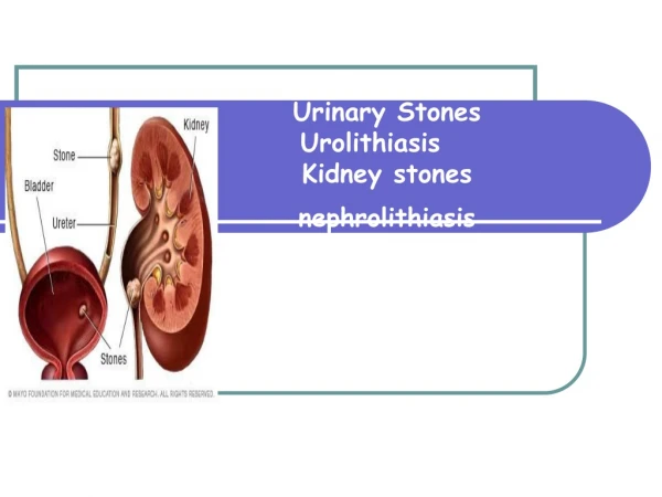
Nephrolithiasis. Eva Jančová. Nephrolithiasis. Renal and ureteral stones are a common problem in primary care practice

E N D
Nephrolithiasis Eva Jančová
Nephrolithiasis • Renal and ureteral stones are a common problem in primary care practice • Primary care physicians need to be alert to the possibility of nephrolithiasis and its consequences to decide upon a diagnostic approach, therapy, and refferal to a urologist or stone specialist
Diagnosis and acute managment of suspected nephrolithiasis • Epidemiology • Etiology • Clinical manifestation • Differential diagnosis • Diagnosis • Acute therapy • Evaluation and subsequent treatment
Diagnosis and acute managment of suspected nephrolithiasis • Epidemiology • Etiology • Clinical manifestation • Differential diagnosis • Diagnosis • Acute therapy • Evaluation and subsequent treatment
Epidemiology • The prevalence of renal calculi varies with the population studied • The rate of nephrolihiasis increases with • age (is higher in men compared to woman) • whites compared to blacks • Patients discharged from hospital with diagnosis of stones • 1.8 per 10 000 • Incidence of stones in general practice • 7 per 10 000 • General practice incidence of stones in males aged 45–60 years • 21 per 10 000
Diagnosis and acute managment of suspected nephrolithiasis • Epidemiology • Etiology • Clinical manifestation • Differential diagnosis • Diagnosis • Acute therapy • Evaluation and subsequent treatment
Etiology • 85% of pts with n. form calcium stones • most of which are composed primary of calcium oxalate • less often calcium phosphate • The other main types include • Uric acid • Struvite (magnesium ammonium phosphate) • Cystine stone • The same pts may have more than one type of stone currently • Calcium and uric acid
Major Risk Factors for Calcium Stones • Low urine volume • Hypercalciuria • Hypocitraturia • Hyperuricosuria • Dietary factors • Low fluid intake • Type of fluid intake-soft drinks, apple or grapefruite juice • High protein intake • Low calcium intake • History of prior calcium stones • Hyperoxaluria (eg, enteric hyperoxaluria) • Meddulary sponge disease • Unexplained association with disorders-hypertension, vasectomy
Other factors affect the risk of stone formation • History of prior calcium nephrolithiasis • Family history of nephrolithiasis • Inhanced of entetric oxalate absorption • Urinary tract infection • Medivations /indinavir, sulfadiazine,…)
Diagnosis and acute managment of suspected nephrolithiasis • Epidemiology • Etiology • Clinical manifestation • Differential diagnosis • Diagnosis • Acute therapy • Evaluation and subsequent treatment
Pain Ureteric colic Lumbar ache On micturition Haematuria Sterile pyuria Asymptomatic proteinuria Dysuria and increased urinary frequency Urinary tract infections Acute (single or recurrent attack) Chronic Pyonephrosis Calculus anuria Strangury and interruption of urine stream Clinical presentations of urinary stones
Patients may occasionally be diagnosed with asymtomatic nephrolithiasis The asymptomatic phase is more likely persist in those who have never had a clinical episode of renal colic Clinical manifestions
Patients occasionally present after already having passed gravel or a stone Uric acid stones are more likely present with gravel but they cal also produce acute obstruction Clinical manifestions-Symptoms
Symptoms are usually produced when stones pass from the renal pelvis into the ureter Pain is the most common symtom and varies from a mild and barely noticeable ache, to discomfort which is so intense that it requires hospitalization and parenteral medications Clinical manifestions
The pain typically waxes and wanes in severity, and develops in waves or paroxysms that are related to movement of the stone in the ureter and associated ureteral spasm Pain is thought to occur due to muscular contraction of the ureter in response to the stone Clinical manifestions-Pain
The site of obstruction determines the locaton of pain Upper ureteral or renal pelvic obstruction lead to flak pain or tenderness Lower ureteral obstruction causes pain that may radiate to the ipsilateral testicle or labia The location of the pain may change as the stone migrates Clinical manifestions-Pain
Variable location of pain can be misleading and occasionally mimics an acute abdomen or dissecting Clinical manifestions-Pain
Gross or microscopic hematuria occur in the majority of patients presenting with symptomatic nephrolithiasis Unilateral flank pain, hematuria, and a positivity plain of the abdomen are present in 90 percent of emergency room patients with a stone Absence of hematuria in the setting of acute flank pain does not exclude the presence of nephrolithiasis Clinical manifestions-Hematuria
Other symptoms Nausea Vommiting Dysuria Urgency Complicantions Persistent renal obstruction Sepsis Clinical manifestions
Diagnosis and acute managment of suspected nephrolithiasis • Epidemiology • Etiology • Clinical manifestation • Differential diagnosis • Diagnosis • Acute therapy • Evaluation and subsequent treatment
Differential diagnosis • Ectopic pregnancy • Aortic aneurysm • Acute intestinal obstruction • Renal carcinoma (bleeding within the kidney)
Diagnosis and acute managment of suspected nephrolithiasis • Epidemiology • Etiology • Clinical manifestation • Differential diagnosis • Diagnosis • Acute therapy • Evaluation and subsequent treatment
Diagnosis • The diagnosis of nephrolithiasis is initially suspected by clinical presentation • Confirmatory radiologic tests include • Abdominal plain film (KUB) • Intravenous pyelography (IVP) • Ultrasonography • CT scan (including spiral CT) • MRI
Abdominal plain film (KUB) Will identify radiopaque stones • Calcium-containing stones • Struvite stones • Cystine stones
Advantages Readily available Inxpensive Limited radiation Useful in acute setting Disadvantages Requies skilled radiologist to interpret Limited sensitivity and specificity Abdominal plain film (KUB)
Intravenous pyelography (IVP) • High sensitivity and specificity for detection of stones and provides data about the degree of obstruction
Advantages Useful in planning therapy and confirming diagnosis Long established history as gold standard Disadvantages Moderately expensive Intravenous contrast required Moderate x-ray exposure Intravenous pyelography (IVP)
Advantages Readily available Roughly equivalent to IVP as a diagnostic test Improved sensitivity with use of color Doppler No radiation exposure Good for hydronephrosis Disadvantages Moderately expensive Poor performance with small stones Requires skilled technician and radiologist Ultrasonography
Advantages Probably new gold standard Can distinguish radiolucent stones from blood and tumor Disadvantages Expensive Moderate x-ray exposure Not uniform available CT scan (including spiral CT)
Advantages Great potential for localizing sight of stone in ureter Disadvantages Vera expensive Largely investigational so far except in certain centres MRI
Results of diagnostic imaging in patients presenting with renal colic Sites of calcific lesions which may be confused with radio-opaque urinary stones: • gallstones, costal cartilages, mesenteric lymph nodes, adrenals, pancreas, renal and splenic arteries, pelvic veins. The radiotranslucent stones • uric acid, xanthine, oxipurinol, 2,8-dihydroxyadenine, orotic acid, and triamterine. Finely stippled nephrocalcinosis suggests • long-standing hypercalcaemia Dense coarse nephrocalcinosis suggests • primary hyperoxaluria or renal tubular acidosis.
Results of diagnostic imaging in patients presenting with renal colic Obstructive uropathy due to: • Radio-opaque stone • Radiotranslucent obstructive lesion (stones, crystals, sloughed papillae, clots, carcinoma) • Generalized nephrocalcinosis • Medullary sponge kidney • Renal papillary necrosis (sloughed papilla) • Cortical scars due to chronic pyelonephritis • Renal carcinoma (cause of ‘clot colic’) • Coincidental calcific lesion (e.g. tuberculosis, Randall’s plaques)
Diagnosis and acute managment of suspected nephrolithiasis • Epidemiology • Etiology • Clinical manifestation • Differential diagnosis • Diagnosis • Acute therapy • Evaluation and subsequent treatment
Acute Therapy • Pain control • Nonsteroidal antiinflammatory drugs (NSAIDs) • Narcotics • Hydration • Urology consultation
Acute Therapy • Patients can be managed at home if they are able to take oral medications and fluids • Hospitalization is required for those who • cannot tolerate oral intake • have very severe pain
Acute Therapy • Hospitalization is required for those who • cannot tolerate oral intake • have very severe pain
Urology consultation Urgent urologic consultation is warranted in patients with • Urosepsis • Acute renal failure Outpatient urology referral is indicated in patients • Who fail to pass the stone after a trial of conservative management (usually two to four weeks) • A stone more 5mm in diameter • Uncontrolled pain
Diagnosis and acute managment of suspected nephrolithiasis • Epidemiology • Etiology • Clinical manifestation • Differential diagnosis • Diagnosis • Acute therapy • Evaluation and subsequent treatment
Evaluation and subsequent treatment • The patient shoud be evaluated for possible underlying causes of stone disease • These include: • Hypercalcemia • Hypercalciuria • Hyperuricosuria • Hypocitraturia • Hyperoxaluria
Evaluation and subsequent treatment-Calcium stones • Composed purely or predominantly of calcium oxalate can occur in many differënt disordes • Calcium phosphate stones are associated with the same risk factors as calcum oxalate stones. One exception- calcium phosphate stones are more typical of complete or incomplete distal renal acidosis
Calcium stones • Oxalate crystals
Urinary risk factors for idiopathic calcium stones • Low volume, which increased the concentrations of the lithogenic factors • Hypercalciuria, with or without hypercalcemia • Hyperuricosuria, which in calcium oxalate-stone formers, is usually due to increased protein intake and therefore uric acid production • Hypocitraturia, which can be marked in patients wih chronic metabolic acidosis • Hyperoxaluria, which may be present in up to 40 percent of male and 15 percent of female stone formers • Defects of macromolecular inhibitors
Urinary risk factors for idiopathic calcium stones • Calcium stone formation is most often idiopathic, but can occur a number of other disorders.
Underlying systemic or renal disorders in calcium stone disease • Primary hyperparathyroidism • Medullary sponge kidney • Distal renal tubular acidosis (complete or incomplete) • Sarcoidosis (and other granulomatous diseases) • Hyperoxaluria • Enteric • Primary
Evaluation and subsequent treatment-Uric acid stones • Pure uric acid stones primary occur in patients in whom a persistently acid urine promotes uric acid precipitation • In the absence of gout, uric acid stones may be seen other causes of chronic overproduction of uric acid or in chronic diarrheal states in which bicarbonate loss and volume depletion lead to a concentrated, acid urine
Caused of Secondary due to increased Purine Biosynthesis and/or Urate Production • Inherited enzyme defects leading to purine overproduction • Clinical disorders leading to purine and/or overproduction • Myeloproliferative disorders • Lymfoproliferative disorders • Malignancies • Hemolytic disorders • Psoriasis • Obesity • Tissue hypoxia • Down Syndrome • Glycogen storage diseases (type III,V,VII)
Caused of Secondary due to increased Purine Biosynthesis and/or Urate Production-continue • Drug-,diet, or toxin-induced purine and /or urate overproduction • Ethanol • Excessive dietary purine ingestion • Pancreatic extract • Fructose • Vitamin B12 (pts with pernicious anemia) • Nicotinic acid • Cytotoxic drugs • Warfarin
Evaluation and subsequent treatment-Struvite stones • Struvite stones only form in pts with a chronic urinary tract infection due to a urease production organism such as Proteus or Klebsiella • The stone may grow rapidly over a period of week to months and, if not, adequately treated, can develop into a staghorn or branched calculus involving the entire renal pelvis and calyces
Evaluation and subsequent treatment-Cystine stones • Cystine stones developt in pts with cystinuria due to the insolubility of cystine in the urine • The diagnosis of cystinuria is made from the family history, by identification of the pathognomonic hexagonal cystine crystal on urinalysis