Download
1 / 81
820 likes | 1.06k Vues
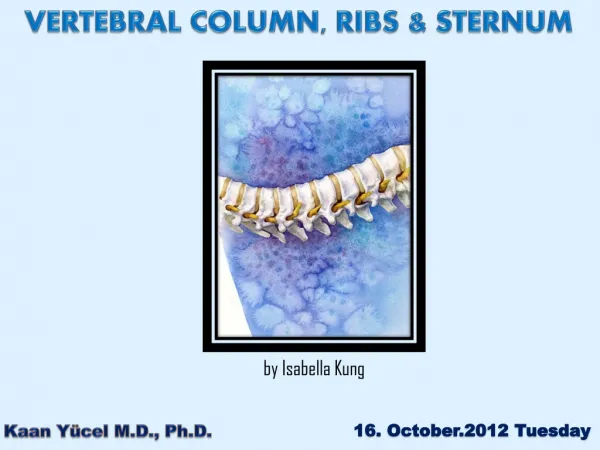
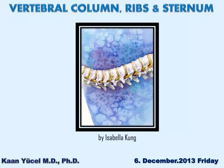
VERTEBRAL COLUMN, RIBS & STERNUM . by Isabella Kung. 6. December.2013 Friday. Kaan Yücel M.D., Ph.D. Vertebrae + intervertebtal (IV) discs Spine Omurga Onurğa Wirbelsäule العمود الفقري Laf dhabar Main part of the axial skeleton. VERTEBRAL COLUMN.

E N D
VERTEBRAL COLUMN, RIBS & STERNUM byIsabella Kung 6. December.2013 Friday Kaan Yücel M.D., Ph.D.
Vertebrae+ intervertebtal (IV) discs Spine Omurga Onurğa Wirbelsäule العمود الفقري Laf dhabar Main part of theaxialskeleton VERTEBRAL COLUMN Mgongo
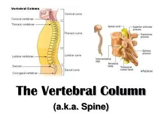
VERTEBRAL COLUMN fromthecranium (skull) totheapex of thecoccyx ¼ formed by the intervertebral (IV) discs. IV discsseparateandbindthevertebraetogether.
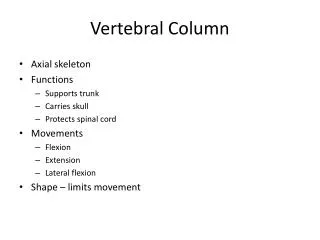
VERTEBRAL COLUMN • Protectsthespinalcordandspinalnerves. • Supportstheweight of the body superiortothelevel of thepelvis. • Providesa partlyrigidandflexibleaxisforthe body and an extendedbase on whichthehead is placedandpivots. • Plays an important rolein postureandlocomotion • (themovementfromoneplacetoanother).
VERTEBRAE • vertebrae(singular = vertebra) • separatedbyresilientintervertebral (IV) discs. • Vertebralcolumnflexible
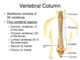
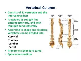
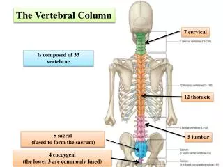
VERTEBRAE 33 vertebraearranged in 5 regions • 7 cervical • 12 thoracic • 5 lumbar • 5 sacral • 4 coccygeal
VERTEBRAE • Significantmotionoccursbetween24 superiorvertebrae. • Of the 9 inferiorvertebrae, 5 sacralvertebraefusedin adultsto form thesacrum • After~ 30, the4 coccygealvertebraefuseto form thecoccyx
VERTEBRAE becomelargeras thevertebralcolumndescendstothesacrum thenbecomeprogressivelysmallertowardapexof thecoccyx
Structures of thevertebrae • A typicalvertebraconsistsof • A Vertebralbody • AVertebralarch • 7 processes 3-4 2 1 5-6 7
VERTEBRAL BODY • Massive, cylndircal • Anteriorpart of the bone • Givesstrength to the vertebralcolumn. • Supportsbody weight. • The size of the vertebral bodiescolumn descends • most markedly from T4 inferiorly • As each bears progressively greater body weight.
Posteriorto the vertebral body • Consists of two (right and left)pedicles& laminae. • VERTEBRAL ARCH
vertebralarch + posterior surface of the vertebral body • walls of vertebral foramen
Successionof vertebral foramina • in the articulated vertebral column • forms • vertebralcanal (spinal canal)
Vertebral notches(Incisura vertebralis) • Indentations observed in lateral views of the vertebrae • Superior and inferior to each pedicle • Between the superiorand inferior articular processes posteriorly • Between the corresponding projections of the body anteriorly.
The superior and inferior vertebral notchesof adjacent vertebrae and the IV discs form intervertebral foramina • Intervertebral foramina • Spinal (posterior root) ganglia are located • Spinal nerves emerge from the vertebral column with their accompanying vessels through these foramina.
Regional Characteristics of Vertebrae • vertebraehaving foramina in their transverse processes are cervicalvertebrae
articularfacetsorientation in eachregiondifferent • Movementneeded • articularfacets of thoracicvertebraenearlyvertical, • define an arc centered in the IV disc • this arrangement permits rotation and lateral flexion of the vertebral column in this region.
Regional variations in size and shape of the vertebral canal accommodate the varying thickness of the spinal cord.
CERVICAL VERTEBRAE skeleton of the neck between the cranium & thoracic vertebrae
FEATURES TYPICAL FOR CERVICAL VERTEBRAE Smallest of the 24 movablevertebrae Relatively larger intervertebral discs discs are thin, but relative to their small size; thick. • 3) Greatest range & variety of movement of all the vertebral regions • 4) foramen transversariumin the transverse process
FEATURES TYPICAL FOR CERVICAL VERTEBRAE 5) anterior tubercles of vertebra C6 carotid tubercles Chassaignactubercles
FEATURES TYPICAL FOR CERVICAL VERTEBRAE 6) Spinousprocesses of C3-C6 short and usually bifid in white people
Vertebrae C3-C7 typicalcervicalvertebrae Largevertebralforamina restrictedrotation superolateralmargin uncusof the body uncinateprocess
C7- vertebraprominens • A longspinousprocess • Mostprominent spinous process in 70% of people
Atlas (C1) No body No spinousprocess Widestof the cervical vertebrae Thekidney-shaped, concavesuperiorarticularsurfaces of thelateralmassesarticulatewithoccipitalcondyles.
Anterior and posteriorarches a tubercle in the center of its externalaspect extendbetweenthelateralmassesforming a complete ring. Posteriorarch A wide groove for the vertebral artery on its superior surface. C1 nerve also runs in this groove.
strongest of the cervicalvertebrae C1, carrying the cranium, rotates on C2 (e.g., when a person turns the head to indicate “no”). Axis (C2)
Axis (C2) The distinguishing featureblunt tooth-like dens Lies anterior to the spinalcord. Serves as the pivot about which the rotation of the head occurs.
Axis (C2) • largebifid spinousprocess
THORACIC VERTEBRAE • The thoracic skeleton includes: • 12 pairs of ribs and associated costal cartilages • 12 thoracic vertebrae and the intervertebral discs between them • Sternum
FEATURES TYPICAL FOR THORACIC VERTEBRAE articulation with ribs. 1) Bilateral costal demifacetson the vertebral bodies for articulation with heads of ribs 2)Costal facets on the transverse processes for articulation with tubercles of ribs
FEATURES TYPICAL FOR THORACIC VERTEBRAE articulation with ribs. 3) Articular processes of thoracic vertebrae extend vertically with paired, nearly coronally oriented articular facets define an arc. greatest degree of rotation is permitted here!
FEATURES TYPICAL FOR THORACIC VERTEBRAE 4)Heart-shaped bodies 5) Long, inferiorly slanting spinous processes
T1-T4 vertebrae share some features of cervical vertebrae. The middle four thoracic vertebrae (T5-T8) demonstrate all the features typical of thoracic vertebrae.
T1atypical 1. long, horizontalspinousprocess Vertebraprominens? No. 2. complete costal facetfor the 1st rib 3. demifacetfor the 2nd rib. Typicalpattern 1+0.5 1+1 costalfacet @ transverseprocesses 0.5+0.5demifacet 0.5+0.5demifacet
[T9]-T10vertebrae No inferiordemifacet 1+1 costalfacet @ transverseprocesses 0.5+0.5demifacet T11-T12vertebrae No transverse costal facets 1 completefacet on eachside 1+1 demifacet
mostcommonlyfracturedvertebra T12 superiorhalfthoracic in character costalfacets& articular processes inferiorhalflumbar in character nocostalfacets articular processes that permit only flexion and extension.
LUMBAR VERTEBRAE in the lower back between the thorax and sacrum
FEATURES TYPICAL FOR LUMBAR VERTEBRAE • massivebodies • transverseprocessesprojectposterosuperiorlyas well as laterally. • mammillaryprocesses & accessoryprocesses
SACRUM L. sacred Wedged-shaped Usually composed of 5 fused sacral vertebrae in adults. Located between the hipbones Sacralcanal continuation of thevertebralcanal in thesacrum.
On the pelvic and posterior surfaces of the sacrum four pairs of sacralforamina
Anterior projecting edge of the body of the S1 vertebra Sacral promontory (L. mountain ridge) importantobstetricallandmark
The sacrum supports the vertebral column and forms the posterior part of the bony pelvis. The sacrum is tilted so that it articulates with the L5 vertebra at the lumbosacral angle. Eur Spine J. 2009 Feb;18(2):212-7. Epub 2008 Nov 18. Assessment of lumbosacral kyphosis in spondylolisthesis: a computer-assisted reliability study of six measurement techniques. Glavas P, Mac-Thiong JM, Parent S, de Guise JA, Labelle H.
The pelvic surface of the sacrum is smooth and concave. • 4 transverselines • Fusion of the sacral vertebrae starts after age20.
The dorsal surface of the sacrum marked by five prominent longitudinal ridges. mediansacralcrest fused rudimentary spinous processes of the superior three or four sacralvertebra
Intermediate sacral crestsfusedarticularprocesses Lateral sacral creststips of the transverse processes of fusedsacralvertebrae
Inverted U-shaped sacral hiatus Sacral cornua (L. Horns) The sacral hiatus leads into the sacral canal. Thesacralcornua, representingtheinferiorarticularprocesses of S5 vertebra, projectinferiorly on eachside of thesacralhiatusandare a helpfulguidetoitslocation.
The superior part of the lateral surface of the sacrum auricularsurface