Download
1 / 39
390 likes | 588 Vues
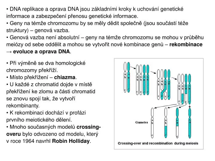
DNA replikace a oprava DNA jsou základními kroky k uchování genetické informace a zabezpečení přenosu genetické informace. Geny na témže chromozomu by se měly dědit společně (jsou součástí téže struktury) – genová vazba.

E N D
DNA replikace a oprava DNA jsou základními kroky k uchování genetické informace a zabezpečení přenosu genetické informace. • Geny na témže chromozomu by se měly dědit společně (jsou součástí téže struktury) – genová vazba. • Genová vazba není absolutní – geny na témže chromozomu se mohou v průběhu meiózy od sebe oddělit a mohou se vytvořit nové kombinace genů – rekombinace → evoluce a oprava DNA. • Při výměně se dva homologické chromozomy překříží. • Místo překřížení – chiazma. • U každé z chromatid dojde v místě překřížení ke zlomu a části chromatid se znovu spojí tak, že vytvoří rekombinanty. • K rekombinaci dochází v profázi prvního meiotického dělení. • Mnoho současných modelů crossing- overu bylo odvozeno od modelu, který v roce 1964 navrhl Robin Holliday.
INITIATION OF RECOMBINATION BY NICKING ONLY ONE PARENTAL MOLECULE The nicked DNA strand invades the other parental molecule by homologous base pairing, thereby displacing a single-stranded loop of DNA. This loop is then cleaved and pairs with the first parental molecule, yielding a crossed-strand Holliday junction. THE HOLLIDAY MODEL FOR HOMOLOGOUS RECOMBINATION Single-strand nicks are introduced at the same position on both parental molecules. The nicked strands then exchange by complementary base pairing, and ligation produces a crossed-strand intermediate called a Holliday junction.
ISOMERIZATION AND RESOLUTION OF HOLLIDAY JUNCTIONS Holliday junctions are resolved by cutting and rejoining of the crossed strands. Two rotations of the crossed-strand molecule, however, produce an isomer in which the unbroken parental strands, rather than the initially nicked strands, are crossed. Cutting and rejoining of the crossed strands of this isomer yield progeny that are recombinant heteroduplexes.
Gene conversion is an event in DNA genetic recombination. It is a process by which DNA sequence information is transferred from one DNA helix (which remains unchanged) to another DNA helix, whose sequence is altered. • Efficient gene conversion generally requires homology between interacting sequences, the homology between the interacting sequences is always >92% and usually >95%.
The double-strand break (DSB); 3′ ssDNA tails; the tails actively ‘scan’ the genome for homologous sequences, and one tail invades the homologous DNA duplex forming a D-loop, which is then extended by DNA synthesis. • Synthesis-dependent strand annealing (SDSA) model: the invading strand and the newly synthesized DNA are displaced from the template and anneal to the other 3′ end of the DSB, leading to the formation of only gene-conversion events. • The other 3′ end of the DSB is captured, and DNA synthesis and ligation of nicks lead to the formation of double HJs. The Bloom syndrome protein (BLM), topoisomerase IIIα and BLM-associated protein BLAP75 act together to remove the double HJs via convergent branch migration (indicated by dotted arrows at both HJs) leading exclusively to gene conversion. In DSBR, the resolution of the double HJs by an HJ resolvase is predicted to generate an equal number of non-crossover and crossover events. • nature reviews | genetics volume 8 | october 2007
Gene-conversion events have been implicated as the molecular cause of an increasing number of human inheriteddiseases. • Pathogenic gene conversion - the transfer of genetic information from non-functional pseudogenes to their closely related functional genes. • Events in which pseudogenes have acted as donors resulted in the functional loss of the acceptor genes through the introduction of frameshifting, aberrant splicing, nonsense mutations, deleterious missense mutations and so on.
Overview of the C4/CYP21 area, showing two RCCX modules as found on most chromosomes. TNXB is the full-size 68 kb gene for tenascin-X, TNXA (shown as a hatched box; also known as XA) is a truncated pseudogene of 5.7 kb that lacks most of the coding sequence of TNXB and has a deletion of 120 bp (indicated by the small triangle) spanning an exon–intron boundary. CYP21A2 (also known as CYP21B) is the active steroid 21-hydroxylase gene; CYP21A1P (shown in black; also known as CYP21A) is a full-size pseudogene containing several deleterious mutations throughout its sequence, including three in-phase stop codons. The C4 genes express variants of the fourth component of complement with different affinities, known as C4A and C4B. The arrangement with C4A in the telomeric and C4B in the centromeric module is common, but the specificity of the C4 genes cannot be determined by means of the restriction sites shown, and many alternative arrangements have been described in the literature. About three-quarters of all RCCX modules are ‘long’ (33 kb in size); the others are ‘short’ (27 kb). The difference depends on the presence or absence of an endogenous retroviral sequence in one of the introns of the C4 gene. The arrows show the orientation of transcription; there is an overlap between the 3′ sections of the oppositely transcribed genes TNXB and CYP21A2, and of TNXA and CYP21A1P, respectively. Bottom: characteristic TaqI and BglII restriction fragments. Top: scale in kb, with the centromeric RCCX duplication boundary at 0. Hum. Mol. Genet. (2002) 11 (21): 2581-2590.
zahrnuje autosomálně recesivní enzymové defekty steroidogeneze v kůře nadledvin s různým biochemickým a klinickým obrazem Kongenitální adrenální hyperplasie (CAH) • DNA diagnostika CAH - analýza genu pro 21-hydroxylázu(zavedena od roku 2003) • Novorozenecký screening deficitu 21-hydroxylázy (zaveden od roku 2007)
Deficit 21-hydroxylázy (21-OHD) • způsoben mutacemi v genu CYP21A2 • CYP21A2 gen a CYP21PA1P pseudogen, lokalizace na chromozómu 6p21.3 • CYP21A2 a CYP21PA1P: každý obsahuje 10 exonů, nukleotidové sekvence jsou v exonech identické z 98% a v intronech z 96% • incidence: v ČR 1: 11 000
Klinické formy 21-OHD • Klasická se solnou poruchou (Salt Wasting) enzymová aktivita: 0% normální aktivity nejčastější mutace: p.Q318X, p.G110VfsX21, klastr E6, p.R356W, chimérní gen a delece genu 2. Prostá virilizující (Simple Virilizing) enzymová aktivita: 1% normální aktivity nejčastější mutace:p.I172N a c.290-13A/C>G 3. Neklasická (Non Classic-late onset) enzymová aktivita: 20-50% normální aktivity nejčastější mutace:p.P30L a p.V281L
3 clinical forms of CAH have been distinguished: • the classical salt-wasting (SW-CAH), • the classical simple virilising (SV-CAH), • the non-classical CAH (NC-CAH). • In addition to having a severe cortisol deficiency, patients with SW-CAH do not synthesise enough aldosteron and therefore, are not able to maintain sodium homeostasis. In the female foetus, the excess of androgen causes variable degrees of external genital virilisation, and consequently, newborn females have genital ambiguity. • The residual activity of 21OH (aldosterone is synthetized) results in SV-CAH with external genital virilisation in females and also results in signs of precocious pseudopuberty which develop by 8 years of age in female and male patients. • NC-CAH is associated with a moderate deficiency in 21OH and manifests in later childhood or adolescence with precocious pseudopuberty and decreased fertility. Patients with NC-CAH secrete aldosterone normally and most of the male patients diagnosed after puberty are entirely asymptomatic
Mutace způsobující 21-OHD 1/ intergenové rekombinace mezi CYP21 genem a CYP21P pseudogenem ~ 95% mikrokonverze · bodové mutace nerovnoměrný crossing-over během meiózy · chimérní CYP21P/CYP21 gen ·delece CYP21 genu · duplikace CYP21 genu
Vznik chimérního CYP21P/CYP21 genu pomocí nerovnoměrného crossing-overu během meiózy • 2/ mutace v CYP21 genu ~5% • nově vzniklébodové mutace
chi-like 5´ 3´ 1 2 4 5 6 7 9 CH-1 3 8 10 Pro30Leu Gly110ValfsX21 Val281Leu Gln318X 236,237,239 c.290-13A/C>G Ile172Asn Arg356Trp Leu307PhefsX6 minisatellite consensus chi-like sequence CH-2 minisatellite consensus minisatellite consensus chi-like sequence CH-3 CH-4 chi-like sequence chi-like sequence CH-5 chi-like sequence CH-6 CH-7 chi-like sequence Types of chimeric CYP21A1P/CYP21A2 genes. The structure of the functional CYP21A2 gene is depicted by white boxes; black boxes represent the nonfunctional CYP21A1P pseudogene. The arrows indicate mutations which exist in CYP21A1P. 236/237/239 depicts mutations p.Ile236Asn, p.Val237Glu, and p.Met239Lys, these mutations always occur together. In CH-4, the CYP21A1P-CYP21A2 junction site is localised inside intron 2 upstream of c.290-13A/C>G. In CH-6, the CYP21A1P-CYP21A2 junction site is localised between mutations c.290-13A/C>G and p.Gly110ValfsX2.
Specific motifs surrounding DNA sequences involved in gene conversion can either promote or inhibit gene-conversion. Such motifs include polypurine and polypyrimidine tracts, palindrome-like sequences, minisatellite sequences, chi and chi-like sequences and sequences that can adopt tetraplex, Z‑DNA, …. • Chi (crossover hotspot instigator) sites had been originally described as cis-acting motifs 5´-GCTGGTGG-3´around which the rate of Rec-promoted recombinations is elevated chi-like 5´ 3´ 1 2 4 5 6 7 9 CH-1 3 8 10 Pro30Leu Gly110ValfsX21 Val281Leu Gln318X 236,237,239 c.290-13A/C>G Ile172Asn Arg356Trp Leu307PhefsX6 minisatellite consensus chi-like sequence CH-2 minisatellite consensus minisatellite consensus CH-3 chi-like sequence CH-4 chi-like sequence chi-like sequence CH-5 chi-like sequence CH-6 CH-7 chi-like sequence
Metodika 21-OHD 1. MLPA - duplikace CYP21 genu - delece CYP21 genu - bodové mutace (p.G110VfsX21; p.I172N; klastr E6; p.Q318X) 2. genově selektivní Long Expand Template PCR - amplifikace CYP21 genu - amplifikace CYP21P/CYP21 chimérního genu 3. sekundární PCR / ACRS - průkaz 11 mutací: p.P30L, c.290-13A/C>G, p.G110VfsX21, p.I172N, klastr E6 (p.I236N, p.V237E, p.M239V), p.V281L, p.Q318X, p.R356W, L306FfsX6 4. PCR-sekvenování
Naše výsledky u 21-OHD DNA analýza provedena u 267nepříbuzných pacientů: diagnóza potvrzena u 241 probandů u 26 pacientů identifikována pouze 1 mutantní alela charakterizováno celkem 30 typů mutantních alel • 60% bodové mutace - 58,6% z CYP21P (24 % c.290-13A/C>G) - 2,4% nově vzniklé mutace • 33,7% chimérní CYP21P/CYP21 geny - 4 typy • 4,9% delece CYP21 genu • 1,0% CYP21 geny nesoucí 2 a více bodových mutací • 0,4% duplikace CYP21 genu (c.290-13A/C>G; p.Q318X)
Kazuistika deficitu 21-hydroxylázy • r. 2003 • žádost o DNA analýzu pro potvrzení diagnózy CAH • u probandky a pro prenatální dg. (matka v 9. týdnu gravidity) • - DNA byla dostupná od obou rodičů a postiženého dítěte: 7letá dívka, v 5 letech věku nástup předčasné puberty, středně virilizovaný zevní genitál, bazální 17-OHP 133 nmol/l • fenotyp: klasická forma bez solné poruchy
Výsledky DNA analýzy rodiny detekce bodové mutace Q318X v HT stavu (8. exonu CYP21 genu) detekce chimérního CYP21P/CYP21 genu v HT stavu • genově selektivní LR PCR • PCR/ACRS - 10 mutací • LR PCR – delece genu PROČ NASTALA NESHODA MEZI GENOTYPEM A FENOTYPEM ??? ?
Rok 2003 - prenatální diagnostika • kultivované AMC • výsledný genotyp plodu odlišný od genotypu probandky ? X biochemické stanovení plodové vody: negativní výsledek !!! (C17-OHP=4,81 nmol/l; N17-OHP=3-17nmol/l)
Po narození syna bylo provedeno ověření genotypu z prenatální dg. – stejný výsledek, dítě však nemělo klinické projevy CAH !!! PROČ TRVÁ V RODINĚ NESHODA MEZI GENOTYPEM A FENOTYPEM ???
r. 2005 - zdokonalení DNA diagnostiky CAH: • sekvenování celé kódující sekvence • detekce bodové mutacep.P459Sv HT stavu na • maternální alele (10. exonu CYP21 genu)
r. 2007 - zdokonalení DNA diagnostiky CAH: 2) MLPA analýza přestaveb 6p21.3 oblasti detekce duplikace CYP21 genu s p.Q318X v HT stavu na maternální alele A/ Otec: B/ Matka:
Familialní hypercholesterolemie (FH) - mutace v genu pro low-density lipoprotein receptor (LDLR)- frekvence heterozygotů (1:500), frekvence homozygotů (1:1000000) Patogenní mutace v LDL receptoru způsobují ztrátu funkčních receptorů pro LDL částice na povrchu jaterních buněk → zvýšení hladiny LDL v plazmě → ateroskleróza a zvýšené riziko koronární srdeční nemoci
V endoplazmatickém retikulu probíhají posttranslační úpravy LDL receptoru, který je následně transportován k buněčné membráně. Na buněčné membráně dohází prostřednictvím specifické interakce s ApoB(apoliproteinB) k tvorbě komplexů LDLR-LDL částice. PCSK9 (proprotein convertase subtilisin-like kexin type 9)je sekretován z hepatocytů a na jejich povrchu se váže LDLR. LDLR (vázaný i nevázaný k PCSK9) je následně shromážděn v záhybech bohatých na klatrin a internalizován za pomocí LDLRAP1 (LDLR adaptor protein). Po endocytóze jsou LDL částice disociovány z LDLR a degradovány v lysozomu. LDLR je recyklován zpátky k buněčné membráně nebo degradován (LDLR vázaný na PCSK9). Garg A, J Clin Endocrinol Metab, 2007
Mechnizmy mající za následek vznik FH: • Defektní struktura a funkce LDL receptoru, • Defektní vazba ApoB/LDLR, • Defektní struktura a funkce LDLRAP1 (LDLR adaptor protein) (narušená endocytóza ApoB/LDLR), • Abnormálně zvýšená aktivita PCSK9 (zvýšený katabolismus LDLR), • Defektní ABCG5/8 (zvýšení intrabuněčné akumulace cholesterolu v hepatocytech).
Figure 3 The LDLR gene Number of unique allelic variants (2009): 1050 Soutar AK and Naoumova RP (2007) Mechanisms of Disease: genetic causes of familial hypercholesterolemia Nat Clin Pract Cardiovasc Med4: 214–225 doi:10.1038/ncpcardio0836
The LDLR gene is localized at 19p13.2, is composed of 18 exons spanning 45 kb, the transcript is 5.3 kb long and encodes a peptide containing 860 amino acids. • LDLR mutations have been reported along the whole length of the gene in FH patients from around the world. • At present, the number of identified unique LDLR allelic variants is over 1000: 65% of the variants are DNA substitutions, 24% small DNA rearrangements (< 100 bp) and 11% large DNA rearrangements (> 100 bp)
PCR/RFLP nejčastější mutace v genu APOB - p.Arg3527Gln • PCR/RFLP pro detekci nejčastějších mutací v genu LDLR - p.Gly592Glu, p.Asp266Glu, and p.Arg416Trp; • PCR a sekvenování exonu 4 genu LDLR(exon s největším výskytem mutací); • MLPA všech exonů genu LDLR; • PCR a sekvenování promotoru a exonů 1, 5, 6, 9, 10, 12, 14 genu LDLR; • DHPLC exonů 2, 3, 7, 8, 11, 13, 15, 16, 17, and 18 genu LDLR, sekvenování vytypovaných exonů Molekulární diagnostika FH Výsledky: 1945 nepříbuzných pacientů s FH - 252 pacientů(13,0%) sAPOB mutací, 406 pacientů (20,9%) s bodovou mutací nebo mutaci malého rozsahu v LDLR, 37 pacientů (1.9%) s mutaci velkého rozsahu v LDLR. V současné době: genotypovací FH čip: detekce 80 typů mutací popsaných u českých FH pacientů a 77 mutací detekovaných s vysokou frekvencí v jiných evropských populacích + MLPA
Genomic characteristics of deletion and duplication breakpoints in the LDLR gene in Czech FH patients
V současné době je známo 117 typů velkých genomových přeuspořádání v LDLR genu : 100 delecí a17 duplikací. • LDLR gen obsahuje 98 Alu repetic, Alurepetice vytváří 65% intronových sekvencí. • Vznik velkých genomových přeuspořádání v LDLR genu je asociován s Alu repeticemi.
45% lidského genomu je tvořeno transposabilními elementy. • Alu repetice vytváří 10% lidského genomu; počet kopií je 1.3 million. Délka Alurepetice je 300 bp; složena ze dvou příbuzných monomerů; do genomu inzerována prostřednictví ssRNA, která je transkribována RNA polymerázou III. 3´-konec Alu dimeruje ukončen oblastí bohatou na A (100 pb), monomery jsou odděleny oblastí bohatou na A, • Distribuce Alurepetic v genomu vytváří podmínky pronon-allelic homologous recombination(NAHR).
Výsledky analýzy 8 genových přeuspořádání u detekovaných u pacientů s FH NAHR, non-allelic homologous recombination– detekováno u 4 delecí a 2 duplikací r.(promoter_ex2del), r.(ex2_6dup), r.(ex3_12del), r.(ex9_14del), r.(ex9_15del), r.(ex16_18dup)] • 6 delecí/duplikací rekombinace mezi dvěma Alu dimery; detekce výrazné sekvenční identity v oblasti zlomu • 2 delece/duplikace rekombinace mezi Alu monomerem a Alu dimerem; detekce sekvenční identity v oblasti zlomu
NHEJ, nonhomologous end joining (zlomy v dsDNA, následuje spojení konců bez sekvenční homologie mezi rekombinovanými úseky DNA) • Delece r.(ex5_10del) – zlom lokalizovaný v intronu 4 je přítomenna konci AluJov antisense orientaci; zlom lokalizovaný v intronu 10 je na konciAluSx1v sense orientaci. • Duplikacer.(ex4_8dup)– zlom lokalizovaný v intronu 3 je přítomen vMER83 repetici; zlom lokalizovaný v intronu 8 je vAluSx1.
Nonallelic homologous recombination-mediated insertion/deletion (NAHR) Nonhomologous end-joining-mediated deletion (NHEJ) NAHR: rectangles depict directly orientated reapeats sharing high homology (97% to 98%), which align at non-allelic and allelic positions and the subsequent recombination causes deletion and duplication. NHEJ: double-strand breaks (DSB) are created between the two sequences with no homology between each other. The NHEJ system modifies and rejoins the two ends, resulting in the deletion of the segment between the two DSBs. Genome Res. 2009 19: 1516-1526