Download
1 / 35
360 likes | 561 Vues
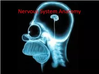
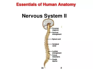
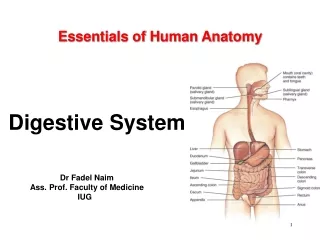
Essentials of Human Anatomy Nervous System I. Chapter 7. Dr Fadel Naim Ass. Prof. Faculty of Medicine IUG. Introduction. The function of the nervous system, along with the endocrine system, is to communicate The nervous system is made up of the brain, the spinal cord, and the nerves.

E N D
Essentials of Human Anatomy Nervous System I Chapter 7 Dr Fadel Naim Ass. Prof. Faculty of Medicine IUG
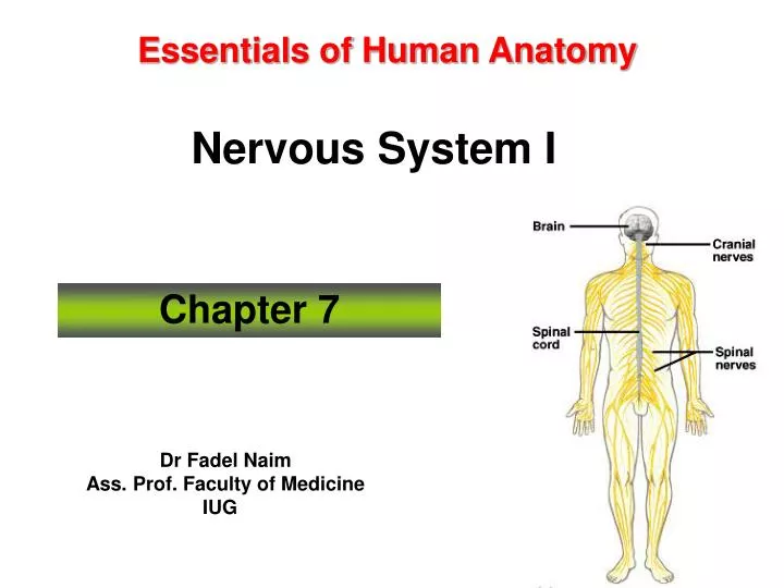
Introduction • The function of the nervous system, along with the endocrine system, is to communicate • The nervous system is made up of the brain, the spinal cord, and the nerves
Functions of Nervous System • Sensory Function • sensory receptors gather information • information is carried to the CNS • Motor Function • decisions are acted upon • impulses are carried to effectors • Integrative Function • sensory information used to create • sensations • memory • thoughts • decisions
Organization of the Nervous System • Organized to detect changes in internal and external environments, evaluate the information, and initiate an appropriate response • Subdivided into smaller “systems” by location: • Central nervous system (CNS) • Structural and functional center of entire nervous system • Consists of the brain and the spinal cord • Integrates sensory information, evaluates it, and initiates an outgoing response • Peripheral nervous system (PNS) • Nerves that lie in “outer regions” of nervous system • Cranial nerves—originate from brain • Spinal nerves—originate from spinal cord
Divisions of Peripheral Nervous System • Sensory Division • picks up sensory information and delivers it to the CNS • Motor Division • carries information to muscles and glands • Divisions of the Motor Division • Somatic – carries information to skeletal muscle • Autonomic – carries information to smooth muscle, cardiac muscle, and glands
Organization of the Nervous System • “Systems” according to the types of organs they innervate • Somatic nervous system (SNS) • Somatic motor division—carries information to the somatic effectors (skeletal muscles) • Somatic sensory division—carries feedback information to somatic integration centers in the CNS
Organization of the Nervous System • Autonomic nervous system (ANS) • Efferent division of ANS—carries information to the autonomic or visceral effectors (smooth and cardiac muscles and glands) • Sympathetic division—prepares the body to deal with immediate threats to the internal environment • Parasympathetic division—coordinates the body’s normal resting activities • Visceral sensory division—carries feedback information to autonomic integrating centers in the CNS
Organization of the Nervous System • Afferent and efferent divisions • Afferent division—consists of all incoming sensory pathways • Efferent division—consists of all outgoing motor pathways
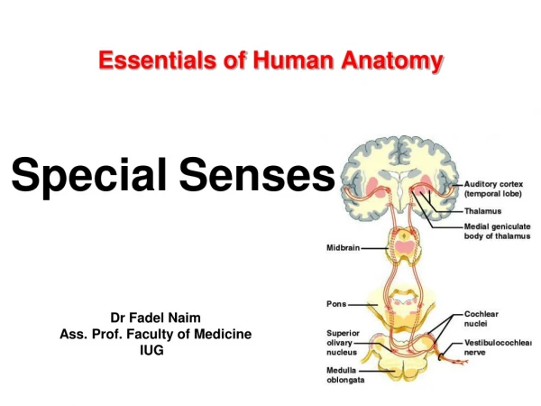
Sensory Division • Somatic sensory components: • General somatic senses: • touch • pain • pressure • vibration, • temperature • proprioception. • Special senses: • Taste • Vision • Hearing • Balance • smell
Sensory Division • Visceral sensorycomponents • transmit nerve impulses from blood vessels and viscera to the CNS • visceral senses primarily include: • temperature • stretch (of the organ wall).
Motor Division • The somatic motor component (somatic nervous system; SNS): • conducts nerve impulses from the CNS to skeletal muscles • also known as the voluntary nervous system • The autonomic motor component (autonomic nervous system; ANS): internal organs, regulates smooth muscle, cardiac muscle, and glands. • Innervates • Internal organs • Regulates smooth muscle • Regulates cardiac muscle • Regulates glands • also known as the visceral motor system or involuntary nervous system
Nerve Cells • Nervous Tissue • Two distinct cell types • Neurons • excitable cells • initiate and transmit nerve impulses • Glial cells • nonexcitable cells • support and protect the neurons
Characteristics of Neurons • Neurons have a high metabolic rate. • Neurons have extreme longevity. • Neurons typically are non-mitotic.
Neuron Structure • Neurons come in all shapes and sizes • All neurons share certain basic structural features. • typical neuron: • Cell body (soma) • Dendrites • Axon
Neuron Structure – Cell Body • The cell body • the neuron’s control center • responsible for: • receiving • integrating • sending nerve impulses. • Consists of: • Plasma membrane • Cytoplasm • Nucleus with prominent nucleolus • Chromatophobic substance (Nissil bodies): RER • Free ribosomes
Cells of the Nervous System • Components of neurons • Dendrites • Each neuron has one or more dendrites, which branch from the cell body • Conduct nerve signals to the cell body of the neuron • Distal ends of dendrites of sensory neurons are receptors
Cells of the Nervous System • Components of neurons • Axon • A single process extending from the axon hillock, sometimes covered by a fatty layer called a myelin sheath • Conducts nerve impulses away from the cell body of the neuron
Neuron Structure – Axon • Structures • Collaterals • Telodendria (axon terminals) • Synaptic knobs (terminal boutons) • The axon transmits a nerve impulse away from the cell body toward another cell.
Classifications of Neurons • Neurons vary widely in morphology and location. • classified based on • structure • function. • Structural classification: number of processes extending from the cell body. • unipolar neuron has a single process • bipolar neurons have two processes • multipolar neurons have three or more processes
Classification of Neurons – Structural Differences • Unipolar • one process • ganglia • Bipolar • two processes • eyes, ears, nose • Multipolar • many processes • most neurons of CNS
Classification of Neurons – Functional Differences • Sensory Neurons • afferent • carry impulse to CNS • most are unipolar • some are bipolar • Interneurons • link neurons • multipolar • in CNS • Motor Neurons • multipolar • carry impulses away from CNS • carry impulses to effectors
Nerves • Nerves are organs of the PNS. • Sensory (afferent) nerves convey sensory information to the CNS. • Motor (efferent) nerves convey motor impulses from the CNS to the muscles and glands. • Mixed nerves: both sensory and motor • Axons terminate as they contact other neurons, muscle cells, or gland cells. • An axon transmits a nerve impulse at a specialized junction with another neuron called synapse.
Peripheral Nerves • Organization – coverings: • Epineurium wraps entire nerve • Perineurium wraps fascicles of tracts • Endoneurium wraps individual axons
Repair of Nerve Fibers • Mature neurons are incapable of cell division; therefore, damage to nervous tissue can be permanent • Neurons have limited capacity to repair themselves • Nerve fibers can be repaired if the damage is not extensive, the cell body and neurilemma are intact, and scarring has not occurred
Regeneration of PNS Axons • PNS axons are vulnerable to cuts and trauma. • A damaged axon can regenerate • if some neurilemma remains. • PNS axon regeneration depends upon three factors. • amount of damage • neurolemmocyte secretion of nerve growth factors • stimulates outgrowth of severed axons • distance between the site of the damaged axon and the effector organ
Repair of Nerve Fibers • Wallerian degeneration: • Stages of repair of an axon in a peripheral motor neuron • Following injury, distal portion of axon and myelin sheath degenerates • Macrophages remove the debris • Remaining neurilemma and endoneurium form a tunnel from the point of injury to the effector • New Schwann cells grow in the tunnel to maintain a path for regrowth of the axon • Cell body reorganizes its Nissl bodies to provide the needed proteins to extend the remaining healthy portion of the axon • Axon “sprouts” appear • When “sprout” reaches tunnel, its growth rate increases • The skeletal muscle cell atrophies until the nervous connection is reestablished • In CNS, similar repair of damaged nerve fibers is unlikely