Download
1 / 1
10 likes | 100 Vues
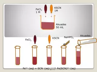
A. B. K. Q. R. HCl 1M 1M 1M 2M 2M 2M 2M temp 20 60 70 70 80 80 100° C min 30 30 30 30 30 60 60. 0M 1M 2M 2M 20 70 80 100° C

E N D
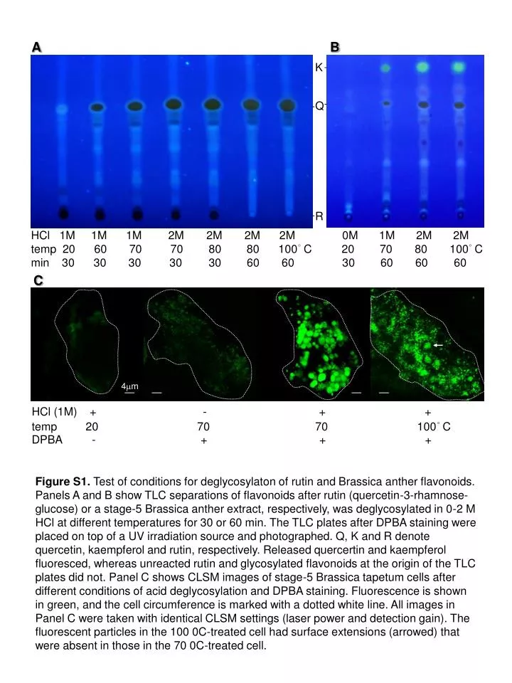
A B K Q R HCl 1M 1M 1M 2M 2M 2M 2M temp 20 60 70 70 80 80 100° C min 30 30 30 30 30 60 60 0M 1M 2M 2M 20 70 80 100° C 30 60 60 60 C 4mm HCl (1M) + - + + temp 20 70 70 100° C DPBA - + + + Figure S1. Test of conditions for deglycosylaton of rutin and Brassica anther flavonoids. Panels A and B show TLC separations of flavonoids after rutin (quercetin-3-rhamnose-glucose) or a stage-5 Brassica anther extract, respectively, was deglycosylated in 0-2 M HCl at different temperatures for 30 or 60 min. The TLC plates after DPBA staining were placed on top of a UV irradiation source and photographed. Q, K and R denote quercetin, kaempferol and rutin, respectively. Released quercertin and kaempferol fluoresced, whereas unreacted rutin and glycosylated flavonoids at the origin of the TLC plates did not. Panel C shows CLSM images of stage-5 Brassica tapetum cells after different conditions of acid deglycosylation and DPBA staining. Fluorescence is shown in green, and the cell circumference is marked with a dotted white line. All images in Panel C were taken with identical CLSM settings (laser power and detection gain). The fluorescent particles in the 100 0C-treated cell had surface extensions (arrowed) that were absent in those in the 70 0C-treated cell.