Download
1 / 12
130 likes | 314 Vues
Assessment of Anterior Chamber Changes after Laser Peripheral Iridotomy using Anterior Segment OCT. Joshua C. Teichman, MD Richard Lee, MD Andrea Butler, BSc Thomas B. Klein, MD FRCSC Iqbal Ike K. Ahmed, MD FRCSC Department of Ophthalmology University of Toronto.

E N D
Assessment of Anterior Chamber Changes after Laser Peripheral Iridotomy using Anterior Segment OCT Joshua C. Teichman, MD Richard Lee, MD Andrea Butler, BSc Thomas B. Klein, MD FRCSC Iqbal Ike K. Ahmed, MD FRCSC Department of Ophthalmology University of Toronto Financial Disclosure: None of the authors have any financial interest in the contents of this poster.
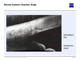
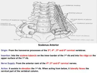
Background • Gonioscopy is the gold standard for evaluating angle anatomy, and the results of laser peripheral iridotomy (LPI) • Subjective, semi-quantitative, affected by pressure and lighting, and difficult to perform • Anterior segment OCT (AS-OCT) may offer a precise, objective, non-contact alternative to gonioscopic evaluation
Purpose • To compare the anatomical changes that occur in the anterior chamber after laser peripheral iridotomy (LPI) using anterior segment optical coherence tomography (AS-OCT)
Methods • Using AS-OCT 74 patients with closed or occludable angles, as determined clinically, were imaged before and after LPI • Low resolution scans of the horizontal and vertical meridians were obtained, as well as high resolution scans of all four quadrants • Scans were conducted in the dark • Patients who had previous surgery that would alter angle anatomy were excluded
Data Measures Anterior chamber depth (ACD) Lens rise (LR:AC) Iris convexity (IC)
Results - Division into Groups • Patients could be divided into two groups • Angles opened significantly after LPI, defined as a change in TIA of > 4° • Angles did not open significantly after LPI, defined as a change in TIA of < 4° • There were 37 patients in each group
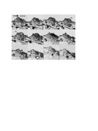
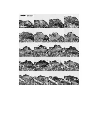
Pre and Post LPI Images Minimal change in angle after LPI Significant change in angle after LPI
Conclusions • In the group of patients whose angles opened significantly after LPI (change in TIA of > 4°): • AOD500increased significantly • ICdecreased significantly • In the group of patients whose angles did not open significantly after LPI (change in TIA of < 4°): • No significant change in AOD500 • IC decreased significantly • This may demonstrate that their narrow angles are likely due to a combined mechanism of pupil block and plateau iris
Conclusions • AS-OCT was useful in the objective measurement of iridocorneal angles before and after LPI • AS-OCT appears to be helpful in differentiating mechanisms of narrow angles: • Pupil block • Lens-related • Plateau iris • AS-OCT may be useful in predicting the effect of LPI preoperatively