Download

1 / 21
240 likes | 446 Vues
Necrotizing enterocolitis Charlene Crichton, MD. Definition. An idiopathic coagulation necrosis and inflammation of the intestine in a neonatal patient Recognized as an important neonatal disorder since the 1960’s. Incidence. The incidence varies from center to center for unknown reasons

E N D
Definition • An idiopathic coagulation necrosis and inflammation of the intestine in a neonatal patient • Recognized as an important neonatal disorder since the 1960’s
Incidence • The incidence varies from center to center for unknown reasons • Affects mostly premature infants (although 10% of cases occur in FT infants) • Increased incidence with decreasing BW and GA with a sharp decrease at 35-36 PCA • Supports the hypothesis that the risk of NEC is determined by maturity of the GI tract
Age of Onset • The age of onset is highly variable but rarely occurs in the first three days of life. • The lowest GA (24-28 weeks) tend to develop NEC after the second week of life • Intermediate GA (29-32 weeks) develop it within 1-3 weeks • Term infants or >32 weeks tend to develop it in the first week of life.
Risk Factors • In the past it was felt that low APGARS, UAC/UVC’s, severe RDS, PDA’s (ie gut ischemia) combined with aggressive and early enteral feeding in a premature infant were the factors associated with NEC • These theories have been dispelled in case-control studies • These studies found that prematurity (with immature GI tract and host defenses) is the primary risk factor
Clinical Manifestations • Bell’s staging criteria Stage I (suspected NEC) Stage II (definite NEC) Stage III (advanced NEC, severely ill) IIIA (without perforation) IIIB (with perforation)
Stage I Systemic signs Intestinal Signs Radiological signs Temp instability, increased A/B’s, lethargy Increased residuals, mild abdominal distention, emesis Normal or mild dilatation or ileus Clinical manifestations
Stage II Systemic signs Intestinal signs Radiologic signs Same as Stage I with metabolic acidosis and mild thrombocytopenia Same as Stage I with decreased bowel sounds and abdominal tenderness Intestinal dilatation, ileus and pneumatosis intestinalis Clinical Manifestations
Stage III (A & B) Systemic signs Intestinal signs Radiologic signs Same as II plus hypotension, severe apnea, DIC, neutropenia, anuria Same as II with generalized peritonitis, marked tenderness and distention, and abdominal wall erythema Same as II with portal vein gas, definite ascites pneumoperitoneum Clinical Manifestations
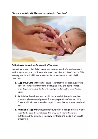
Treatment strategies • Suspected NEC (Bell’s stage I) • Hold enteral feeds • Obtain an x-ray to view bowel gas pattern • Gastric decompression with an OG tube to suction • ROS with initiation of IV antibiotics
Treatment Strategies • Definite NEC (Bell’s stage II) Follow serial exams and serial xray’s with left lateral decubitus films to screen for perforation • Frequent labs with correction of metabolic disturbances(acidosis, hyperkalemia, hyperglycemia etc), hypovolemia, thrombocytopenia, and DIC • Intubation if patient is not on MV • Consider surgical consult
Treatment Strategies • Advanced NEC (Bell’s Stage III) Same management as Stage II with increased monitoring of BP, DIC panels and abdominal films (q6h flat and left lateral decub or cross table lateral films is typical) • Vigorous fluid resuscitation, inotropes, ventilator support • Surgery as indicated
Treatment Strategies • When is surgery indicated?? Absolute indications 1) pneumoperitoneum 2) intestinal gangrene (if the patient is extremely unstable some surgeon opt for peritoneal drains as a bridge to surgery) • Relative indications 1) progressive clinical deterioration 2) fixed abdominal mass, portal vein gas, abdominal wall erythema 3) persistently dilated bowel loop
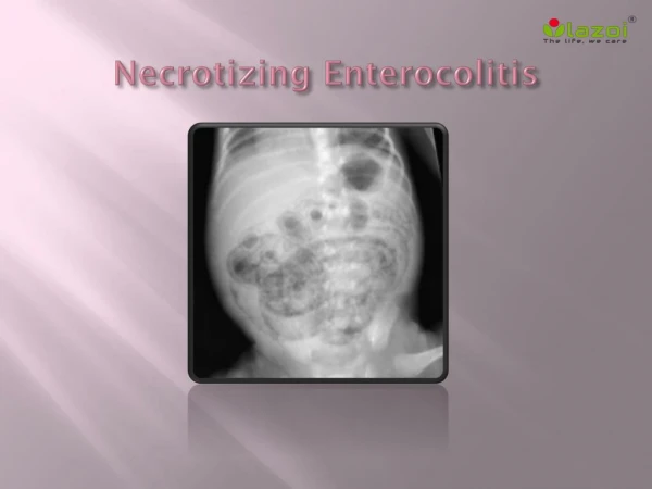
Radiologic findings • Generalized bowel distention (earliest sign) • Pneumatosis Intestinalis • Pneumoperitoneum • Large distended immobile loop on repeated x-rays (persistant loop sign) (may indicate a gangrenous loop of bowel) • Gasless abdomen (perforation and peritonitis) • Portal venous air
Complications • Mortality is 30-60% • Stricture formation is 25-35% • Bowel obstruction in 5% • Enterocutaneous fistulas • FTT secondary to short bowel syndrome and malabsorption • TPN related cholestasis • Central line sepsis
Prevention • Antenatal steroids decreased the incidence of NEC in randomized blinded studies • Use of human milk (1.2% incidence vs. 7.2% incidence in formula feed premies) • GI priming with cautious advancement of enteral feeding