Download
1 / 1
20 likes | 149 Vues
Alterations in Acute and Late Cytokine Expression Correlate with Radiation-induced Fibrosis Simone CB II 1 , Ly D 1 , Soule BP 2 , Savage JE 1 , Mitchell JB 2 , Simone NL 1

E N D
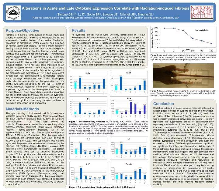
Alterations in Acute and Late Cytokine Expression Correlate with Radiation-induced Fibrosis Simone CB II1, Ly D1, Soule BP2, Savage JE1, Mitchell JB2, Simone NL1 National Institutes of Health, National Cancer Institute, 1Radiation Oncology Branch and 2Radiation Biology Branch, Bethesda, MD Purpose/Objective Fibrosis is a normal consequence of tissue injury and chronic inflammation. Fibrosis is characterized by the accumulation and activation of excessive fibroblasts, deposition of extracellular matrix proteins, and distortion of normal tissue architecture. External beam radiation therapy induces both acute and late fibrotic changes in normal tissues that can lead to loss of normal tissue function and significant patient morbidity. Transforming growth factor (TGF)-β is considered to be a primary inducer of tissue fibrosis, and it has previously been demonstrated to play a role specifically in radiation-induced fibrosis. IL-13 has also been implicated as an inducer of tissue fibrosis. The effects of IL-13 were initially believed to be related solely to its regulation of the production and activation of TGF-β, but more recent investigation has demonstrated IL-13-mediated fibrosis via a pathways independent from TGF-β. Eosinophils might also be responsible for the production of pro-fibrotic cytokines, including IL-13 and TGF-β. Little is known, however, regarding which other cytokines are important regulators in the development of acute or chronic fibrosis. Even fewer data is available regarding the effects that radiation therapy has on these cytokines. This in vivo study examines the acute and late effects of radiation on cytokines previously reported to have a qualitative association with fibrogenesis. Materials/Methods The right hind legs of nine-week old CH3 mice were irradiated in a single 35 Gy fraction. Mice were sacrificed at 1 hour, 7 days, 14 days, 30 days, 90 days, or 120 days post-irradiation. Left hind legs served as controls. Protein was extracted by homogenizing with a protease inhibitor containing tissue protein extraction reagent (Thermo-scientific, Rockford, IL) in an approximately 1:20 W/V ratio. The samples were spun at 4ºC at 10,000 g for 10 minutes. After the supernatant was isolated, the samples were then spun at 4ºC at 10,000 g for 5 minutes. The supernatant was isolated again and the protein concentration was assessed by the Bio-Rad DC Protein Assay (Bio-Rad, Hercules, CA) according to the manufacturer instructions. The protein lysate samples were sent to Quansys Biosciences (Logan, UT) for cytokine and chemokine analysis of IL-1α, IL-1β, IL-2, IL-4, IL-5, IL-6, IL-10, IL-17, MCP-1α, IFN-γ, MIP-1α, TNF-α, Eotaxin, GM-CSF, and CXCL-1 using a custom multiplex ELISA that allowed for the testing of multiple cytokines and chemokines in each well. IL-13 and TGF-β were evaluated individually by single analyte ELISAs according to the manufacturer instructions (R&D Systems, Minneapolis, MN). All sampled were run in triplicate of a three-step dilution. Expression of each cytokine was compared to controls for each time point and normalized according to protein concentration. Conclusion Radiation induced an acute cytokine response reflected in a near global increase in cytokine expression 1 hour post-irradiation most evident for IL-13 (894.4%) and IL-6 (412.6%). Subacutely (days 7, 14, 30), cytokine expression was generally decreased below baseline levels. This may represent the suppressive effects of anti-inflammatory regulatory mechanisms. In the late setting (days 90, 120), increased expression of TH17-mediated and other pro-inflammatory cytokines (IL-1α, IL-1β, IL-6, TGF-β) and TH2/eosinophil-associated pro-fibrotic cytokines (IL-4, IL-5, IL-13, Eotaxin) was observed. This coincided with a decrease in anti-inflammatory IL-10. Radiation appears to induce dysregulated cytokine production and alter the expression of both TH2/eosinophil-associated cytokines and cytokines that influence inflammation. While each of these pathways has been implicated in tissue fibrogenesis independently, these data suggest that both pathways may mediate radiation-induced fibrosis in both the acute and late settings. Radiation-induced fibrosis may, in part, be eosinophilic mediated. Activation and recruitment of eosinophils, which are highly dependent on IL-5, may result in an amplification of fibrotic change or may be a strong mediator of fibrogenesis by upregulating pro-fibrotic cytokines, such as IL-13 and TGF-β, that serve as the main mediators of tissue fibrosis. Therapies that modulate cytokine expression, such as targeted immunomodulators, may alter the development or progression of radiation-induced fibrosis and may improve radiation therapy morbidity. Figure 1. Pro-inflammatory cytokines (IL-1α, IL-1β, IL-6, TGF-β). Mean cytokine expression normalized to the cytokine concentration of unirradiated control samples at time 0, set as a reference of 100.00%. The relative percentage difference is depicted for samples 1 hour, 7 days, 14 days, 30 days, 90 days, and 120 days following radiation, with radiated sample values adjusted to control sample values at each corresponding time point. All sampled were run in triplicate of a three-step dilution. Figure 2. TH2-associated pro-fibrotic cytokines (IL-4, IL-5, IL-13, Eotaxin). Results All cytokines except TGF-βwere uniformly upregulated at 1 hour following radiation when compared to controls (range 9.0% to 894.4%). Cytokines were all downregulated 7, 14, and 30 days following radiation, with the exceptions of TGF-β (upregulated 239.8% at day 14, 169.2% at day 30), IL-13 (162.5% at day 7, 30.7% at day 30), and Eotaxin (76.5% at day 30). At day 90, radiated samples showed moderate upregulation of IL-1β, IL-4, and IL-10 (range 11.0% to 23.6%), and significant upregulation of IL-5, IL-6, MIP-1α, Eotaxin, and CXCL-1 at day 90 (range 44.7% to 123.3%). Of cytokines upregulated by radiation at day 90, only IL-1β, IL-5, and IL-6 remained upregulated at day 120 (range 19.0% to 106.0%). Irradiated IL-13 (165.1%), TGF-β (140.0%), and IL-1α (55.0%) were also significantly upregulated at day 120 (Figures 1-2). Leg Length Ratio Days Figure 3. Leg length ratio. Mean ratio of the length of the right hind leg to the length of the left hind leg of CH3 mice by time in days from irradiation to the right hind leg expressed as a percentage change from the control. Figure 4. Representative image depicting the length of the hind legs of CH3 mice. The right hind leg was irradiated 120 days earlier with a single 35 Gy fraction. The left hind leg serves as a control.