Download
1 / 38
410 likes | 861 Vues
Morphological Changes of Liposomes Induced by Melittin. Jirun Sun Full Talk for Macromolecules Seminar. J, Dufourcq et al., BBA 859, 1986, 33. Contents. Background Introduction of Liposomes Introduction of Melittin Morphological changes of Liposomes with Melittin Position of Melittin

E N D
Morphological Changes of Liposomes Induced by Melittin Jirun Sun Full Talk for Macromolecules Seminar J, Dufourcq et al., BBA 859, 1986, 33
Contents • Background • Introduction of Liposomes • Introduction of Melittin • Morphological changes of Liposomes with Melittin • Position of Melittin • Pore Formation in Liposomes • Morphological Changes of Liposomes
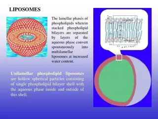
What Are Liposomes • Artificial membrane vesicles. • Drug delivery, chemical manufacturing , genetic engineering and so on http://www.ncnr.nist.gov/programs/reflect/rp/biology/cell_membrane.html
Each phospholipid includes • a polar region: glycerol, carbonyl of fatty acids, Pi, & the polar head group (X) • 2 non-polar hydrocarbon tails of fatty acids (R1, R2). Such an amphipathic lipid may be represented as at right. 3D picture of a Phospholipid
According to the head group X, there are several kinds of lipid. X= Serine (PS) Choline (PC) Inositol (PI) O-H Acid(PA) Glycerol (PG) Ethanolamine (PE)
The membrane lipid composition in an average mammalian cell Phosphatidylcholine (PC), or lecithin, with choline as polar head group. It is a common membrane lipid.
Double bonds in fatty acids usually have the cis configuration. Most naturally occurring fatty acids have an even number of carbon atoms. Some fatty acids and their common names: 14:0 myristic acid (DM); 16:0 palmitic acid (DP); 18:0 stearic acid (DS); 18:1 cisD9 oleic acid (DO) DOPC: two oleic acid tails and a Choline head group POPA: one P tail and one O tail, Acid head group
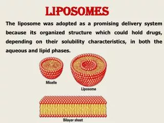
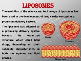
Liposomes Formed by Lipids Bilayer Micelle Driven by hydrophilic and hydrophobic forces, the nonpolar tails of lipids (U) tend to cluster together, forming a lipid bilayer (1), a micelle (2). The polar heads (P) face the aqueous environment, Liposomes contain bilayers. http://www.nupedia.com/newsystem/upload_file/830/bilayer_micelle.png
Size Determined by Methods MLV: Multilamellar vesicles SUV: Small unilamellar vesicles LUV: Large unilamellar vesicles GUV:Giant unilamellar vesicles Sonication: SUV Smaller than 100 nm diameter Extrusion: LUV (Size depends on the filters) 100 nm—1 µm diameter Evaporation: GUV Larger than 1 µm diameter http://www.avantilipids.com/PreparationOfLiposomes3Big.html
Methods to Check the Morphology of liposomes • Freeze-fracture electron microscope (FFEM) or electron microscope (EM) • Light scattering (LS)
Freeze-fracture electron Micrographs (a, b, c) and negative staining (d) of aqueous dispersions of DOPC/DOPA (80:20 mol%) after: 0(a); 10 (b and d) and 50 (c) cycles of freeze-thawing Eur Biophys J 2000 29; 184
Index-matching liquid Incident beam qi Test tube θ qs Unscattered beam q qi Photodetector Static Light Scattering Liposomes: DOPC 14 angles are checked Top view of the geometry around the sample cell Rg= 519±3 Å
Dynamic Light Scattering (DLS) Liposomes: DOPC Six angles, 30,40,50,60,70 and 90 degree were checked. Intensity Measured by Autocorrelation function Rh= 500±7 Å G(2)(t) = <I(0)I(t)> = The signal in the correlator is well approximated by G(2)(t) = B(1 + fg(1)(t)2) g(1)(t) is a simple exponential g(1)(t) = e-Gt Stokes’ law
What do SLS and DLS tell us? If Rg/Rh ≈ 1 the thickness of a ball is about zero and then it more like a thin bubble. For liposomes, they are Unilamellar Rh Rg According to the DLS and SLS data in the previous two slides Rg= 519±3 Å Rh= 500±7 Å Rg/Rh= 1.04 That DOPC is Unilamellar
Phase Transition Temperature (Tm) • Important when preparing the liposomes • Important in the interactions of Melittin with liposomes http://www.virtuallaboratory.net/Biofundamentals/lectureNotes/Topic2-3_Membranes.htm
Melittin • It is the main component (50-60%) in bee venom with 26 residues • It has a very strong anti-inflammatory and anti-bacterial effect • It may cause allergic reaction, which can act as a skin, eye or respiratory irritant. • It is a cytolytic protein. • It is an amphiphile protein. http://molvis.chem.indiana.edu/C581_F97/protein_projects/Melittin.html Tervilliger et.al., Nature, 1982, (299),371
The fact that melittin can act as a lytic agent is partly due to its detergent-like sequence. Hydrophilic Hydrophobic ∂ -helical wheel of the first 20 residues. Leippe et al. PNAS, 88 (1991) 7659
Morphological Changes of Liposomes with Melittin • A weapon of bees • The most popular model for studying cytolytic activities • Membrane fusion • Drug releasing system • Well understanding of the interaction will be helpful on • Building new instruments • And studying new amphiphile proteins
Points • Melittin position in liposomes • The existence of poles • Morphology changes of liposomes • MLV or unilamellar • Ratio of lipid to melittin • Temperature • Time
Melittin in Membranes • Tetrameric aggregate • End to end distance of melittin from crystal structure is about 3.6 nm Simulation of melittin tetrameric aggregate The N-terminus and the C-terminus are denoted By N and C, respectively. The four helices are Labeled 1-4. Lin, et. al., Biophysical Journal, 78, 2000, 1714 Tervilliger et.al., Nature, 299, 1982, 371
Models of Pores Toroidal model fits melittin better Barrel-stave model The difference is whether the water core is lined by both the peptides and the lipid head groups Toroidal model Biophysical Journal, 81, 2001, 1475
Figures of Molecular Dynamics Simulation by Monte Carlo program Snapshot of the pore from top (A) and from side (B). Lin, et. al., Biophysical Journal, 78, 2000, 1714
Existence of Poles • Prepare liposomes in the presence of fluorescent marker, like calcein • Remove external calcein • Measure background, set calcein releasing as zero • Add melittin • Measure the released calcein by spectrofluorimeter.
Leakage Experiments Permeabilzing capacity of melittin on MLV. The release of calcein from POPC/SMPC = 2:1 (mol/mol). Melittin Concentration 1 µm. G. Anderluh et. al., JBC, 278, 2003, 45216
Morphology Changes of Liposomes • Temperature effects (Gel Phase or liquid crystal phase) • Unilamellar or multi-lamellar • Ratio of lipid to melittin matters • It is a dynamic process. • Minor factors like buffer, pH values and so on • That are combining results of all factors.
Melittin is Detergent-Like But They Are Not the Same Fusion happens! Note for the cartoon: DPPC LUV L:M = 200 L:D = 200 Incubation: heat to T>Tm and hold there for a while and then cool down and measure at T<Tm. No Fusion C. G. Morgan et. al., BBA 732 (1983)668
Morphology Changes at Different Ratios Temp: 21 oC Rh by checked by Quasi-elastic LS EPC: Egg PC EPC is in LC state at 21 oC Data digitalized according to: J, Dufourcq et al., BBA 859, 1986, 33
Different L/M Ratios of EPC SUV Seen by FFEM A: Pure EPC SUV B: L:M = 200 C: L:M = 30 D: L:M = 5 Magnification: 50,000x From J, Dufourcq et al., BBA 859, 1986, 33
Different L/M Ratios of EPC MLV Seen by FFEM A: Pure EPC MLV B: L:M = 30 C: L:M = 15 Magnification: 50,000x From J, Dufourcq et al., BBA 859, 1986, 33
The Different Between MLV and SUV MLV + Melittin Vesicularisation Fragmentation LUV+ Melittin Tiny particles Fusion SUV + Melittin J, Dufourcq et al., BBA 859, 1986, 33
Gel Phase or LC Phase LC phase: Morphology changes, Melittin binds to vesicles Gel phase: Only SUVs are morphologically affected Incubation: Morphology changes It is not reversible!
Morphological Changes at Different Temperatures Large particles scatter more! Liposome used DPPC MLV, Ratio of liposomes to melittin L:M=20 Tm for DPPC is 41 oC Data digitalized according to: J, Dufourcq et al., BBA 859, 1986, 33
Morphological Changes Detected by Freeze-fracture EM DPPC MLV DPPC: melittin = 30 A: temp 20 oC B: temp 50 oC C: temp 20 oC after incubation at 50 oC From J, Dufourcq et al., BBA 859, 1986, 33
It Is A Dynamic Process Size changing of liposome (DOPC LUV) by adding melittin checked by Rapid SLS using a commercially available, 18-detector instrument One of two “Delta Dawns” at LSU • Procedure: • Measure background • Normalize with Tol or Emulsion • Measure sample • Data processing A touch is needed! Data from Dr Russo’s Lab agree with the results in literature: J, Dufourcq et al., BBA 859, 1986, 33
Conclusion • Liquid crystal state has more obvious morphology changes than gel state does. • The ratio of L/M matters • MLV, SUV behave differently with melittin • It is a dynamic process