Download
1 / 37
750 likes | 3.24k Vues
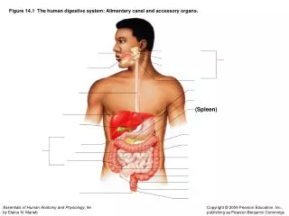
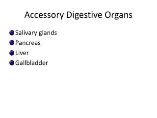
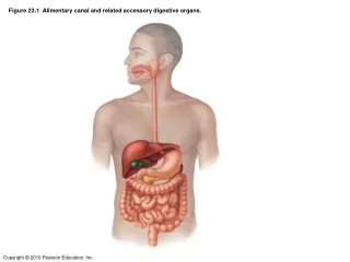
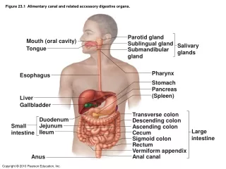
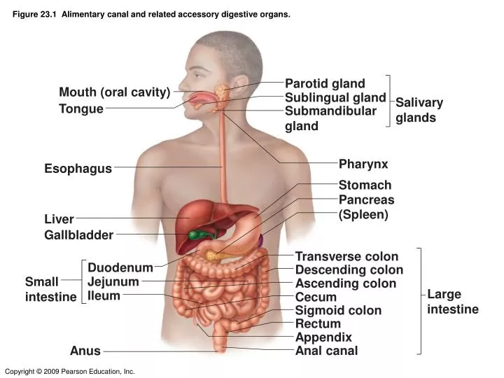
Figure 23.1 Alimentary canal and related accessory digestive organs. Parotid gland. Mouth (oral cavity). Sublingual gland. Salivary glands. Tongue. Submandibular gland. Pharynx. Esophagus. Stomach. Pancreas. (Spleen). Liver. Gallbladder. Transverse colon. Duodenum.

E N D
Figure 23.1 Alimentary canal and related accessory digestive organs. Parotid gland Mouth (oral cavity) Sublingual gland Salivary glands Tongue Submandibular gland Pharynx Esophagus Stomach Pancreas (Spleen) Liver Gallbladder Transverse colon Duodenum Descending colon Jejunum Small intestine Ascending colon Large intestine Ileum Cecum Sigmoid colon Rectum Appendix Anal canal Anus
Figure 23.1 Alimentary canal and related accessory digestive organs.
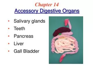
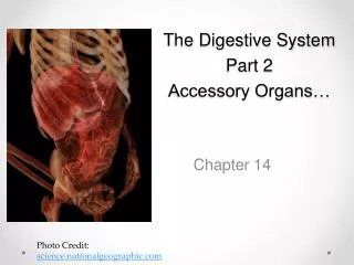
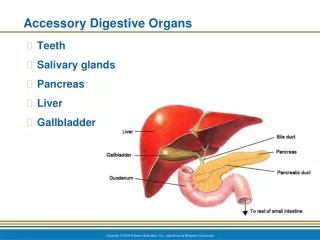
Figure 14.1 Organs and accessory organs of the digestive system and their functions.
Figure 23.6 Basic structure of the alimentary canal. Glands in submucosa Mucosa • Epithelium • Lamina propria • Muscularis mucosae Submucosa Muscularis externa • Longitudinal muscle • Circular muscle Serosa Nerve • Epithelium Artery • Connective tissue Vein Lumen Gland in mucosa Lymphatic vessel Mucosa-associated lymphoid tissue Duct of gland outside mucosa Mesentery
Figure 23.7b Anatomy of the oral cavity (mouth). Gingivae (gums) Hard palate Soft palate Uvula Palatine tonsil Tongue Sublingual fold with openings of sublingual ducts Opening of submandibular duct
Figure 23.9a The salivary glands. Ducts of sublingual gland Tongue Teeth Parotid gland Parotid duct Body of mandible (cut) Sublingual gland Submandibular duct Submandibular gland (a)
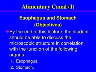
Esophagus • Mucosal epithelium is stratified squamous Stomach • Mucosal epithelium is simple columnar
Liq uified food in the intestinal tract, first produced in the stomach is chyme. • Contents of the intestinal tract in the fetus is meconium.
Figure 23.14a Anatomy of the stomach. Esophagus Muscularis externa Serosa • Longitudinal layer • Circular layer Body • Oblique layer Lumen Lesser curvature Rugae of mucosa Greater curvature Duodenum Pyloric sphincter (valve) at pylorus (a)
Figure 23.16 Photographs of a gastric ulcer lesion and of the bacteria that most commonly cause it. Bacteria Mucosa layer of stomach (b) H. pylori bacteria (a) A gastric ulcer lesion
Microscopic Anatomy of the Stomach Figure 22.15
Small Intestine • Three subdivisions: duodenum, jejunum, and ileum • The bile duct and main pancreatic duct join the duodenum at the hepatopancreatic ampulla
Figure 14.10 Locations and digestive functions of the liver, gallbladder, and pancreas.
Figure 14.10 Locations and digestive functions of the liver, gallbladder, and pancreas. 4 1 2 6 8 5 7 3
Figure 23.21 The duodenum of the small intestine, and related organs. hepatic ducts of liver Bile duct Pancreas Gallbladder Jejunum Main pancreatic duct Hepatopancreatic ampulla and sphincter Duodenum
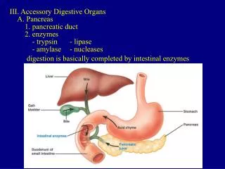
Pancreas • Exocrine function • Secretes pancreatic juice which contains enzymes which break down all categories of foods • Secretes HCO3– which neutralizes acidic chyme, and provides optimal environment for pancreatic enzymes • Enzymes are released in inactive form and activated in the duodenum
Figure 23.26a Structure of the enzyme-producing tissue of the pancreas. Small duct Acinar cells Basement membrane Zymogen granules Rough endoplasmic reticulum (a)
Liver • The largest gland in the body • Has four lobes • Hepatic artery- brings oxygen rich blood to the liver • Hepatic portal vein- brings nutrient rich blood from the digestive organs to the liver • Both of these empty into the sinusoids of the liver
Figure 23.25a-b Microscopic anatomy of the liver. (a) (b) Lobule Central vein Connective tissue septum
Liver: Microscopic Anatomy • Liver sinusoids – enlarged, leaky capillaries • Kupffer cells – hepatic macrophages found in liver sinusoids • Hepatocytes’ functions include: • Production of bile • Processing bloodborne nutrients • Storage of fat-soluble vitamins • Detoxification • Protein synthesis • Synthesis of cholesterol • Secreted bile flows between hepatocytes toward the bile ducts
Figure 23.25c Microscopic anatomy of the liver. Interlobular veins (to hepatic vein) Central vein Sinusoids Bile canaliculi Plates of hepatocytes Bile duct (receives bile from bile canaliculi) Bile duct Portal venule Portal triad Hepatic macrophages in sinusoid walls Arteriole Portal vein (c)
Large Intestine • Subdivided into the cecum, appendix, colon, rectum, and anal canal • Cecum and appendix have digestive function in herbivores • Has three bands of longitudinal smooth muscle in its muscularis
Figure 23.30d Mesenteries of the abdominal digestive organs. Liver Lesser omentum Pancreas Stomach Duodenum Transverse colon Mesentery Greater omentum Jejunum Ileum Visceral peritoneum Parietal peritoneum Urinary bladder Rectum (d)
Figure 23.30a Mesenteries of the abdominal digestive organs. Liver Gallbladder Spleen Stomach Greater omentum Small intestine Cecum (a)
Figure 23.30b Mesenteries of the abdominal digestive organs. Liver Gallbladder Lesser omentum Stomach Duodenum Transverse colon Small intestine Cecum (b)
Figure 23.30c Mesenteries of the abdominal digestive organs. Transverse colon Descending colon Jejunum Mesentery Sigmoid colon Ileum (c)
Tooth Structure Figure 22.11
Two main regions – crown and the root Crown – exposed part of the tooth above the gingiva (gum) Covered with enamal, the hardest substance in the body, which is composed of calcium and phosphate salts Root – portion of the tooth embedded in the jawbone Covered with cementum, which is also calcified Tooth Structure 1
Periodontal ligament Anchors the tooth in the jaw Dentin – bonelike material beneath the enamel cap that forms the bulk of the tooth Pulp cavity –center of tooth, containing connective tissue, blood vessels, and nerves Root canal – extension of the pulp cavity out of the root Tooth Structure 2
Teeth are classified according to their shape and function Incisors: chisel-shaped teeth adapted for cutting or nipping Canines: conical or fanglike teeth that tear or pierce Premolars (bicuspids) & molars (tricuspids): have broad crowns with rounded tips and are best suited for grinding or crushing
Figure 23.10a Human dentition. (DON’T NEED TO KNOW AGE OF APPEARRANCE OF TEETH) Incisors Incisors Central (7 yr) Central (6–8 mo) Lateral (8 yr) Lateral (8–10 mo) Canine (eyetooth) (11 yr) Canine (eyetooth) (16–20 mo) Premolars (bicuspids) Molars First molar (10–15 mo) First premolar (11 yr) Deciduous (milk) teeth Second molar (about 2 yr) Second premolar (12–13 yr) Molars First molar (6–7 yr) Second molar (12–13 yr) Third molar (wisdom tooth) (17–25 yr) Permanent teeth (a)