Download
1 / 26
260 likes | 500 Vues
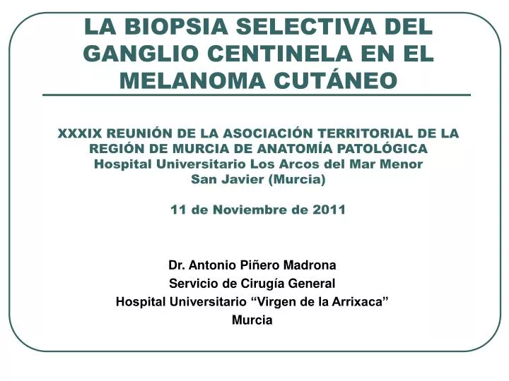
LA BIOPSIA SELECTIVA DEL GANGLIO CENTINELA EN EL MELANOMA CUTÁNEO XXXIX REUNIÓN DE LA ASOCIACIÓN TERRITORIAL DE LA REGIÓN DE MURCIA DE ANATOMÍA PATOLÓGICA Hospital Universitario Los Arcos del Mar Menor San Javier (Murcia) 11 de Noviembre de 2011. Dr. Antonio Piñero Madrona

E N D
LA BIOPSIA SELECTIVA DEL GANGLIO CENTINELA EN EL MELANOMA CUTÁNEOXXXIX REUNIÓN DE LA ASOCIACIÓN TERRITORIAL DE LA REGIÓN DE MURCIA DE ANATOMÍA PATOLÓGICAHospital Universitario Los Arcos del Mar MenorSan Javier (Murcia)11 de Noviembre de 2011 Dr. Antonio Piñero Madrona Servicio de Cirugía General Hospital Universitario “Virgen de la Arrixaca” Murcia
CONCEPTOS GENERALES • Importancia de conocer la afectación ganglionar: • Estadificación … TNM • Tratamiento locorregional (en casos de afectación establecida). • Métodos para determinar la afectación ganglionar: • Exploración clínica. • Conservadores (métodos de imagen): • Ecografía. • RMN. • PET-TC. • Invasivos: • Linfadenectomía. • BSGC.
LINFADENECTOMÍA ELECTIVA • No importancia pronóstica: • Veronessi U et al. Delayed regional lymph node dissection in stage I melanoma of the skin of the lower extremities. Cancer 1982; 49: 2420-2430. • Sim FH et al. Lymphadenectomy in the management of stage I melanoma: a prospective randomized study. Mayo Clin Proc 1986; 61: 697-705. • Balch M et al. Efficacy of an elective regional lymph node dissection of 1-4 mm thick melanomas for patients 60 year of age or younger. AnnSurg 1996; 224: 255-266. • Cascinelli N et al. Immediate or delayed dissection of regional nodes in patients with melanoma of the trunk: a randomized trial. Lancet 1998; 351: 793-796. • Dificultad para determinar drenajes específicos: • Drenajes no esperados. • Drenajes múltiples.
LINFADENECTOMÍA ELECTIVA • No importancia pronóstica: • Veronessi U et al. Delayed regional lymph node dissection in stage I melanoma of the skin of the lower extremities. Cancer 1982; 49: 2420-2430. • Sim FH et al. Lymphadenectomy in the management of stage I melanoma: a prospective randomized study. Mayo Clin Proc 1986; 61: 697-705. • Balch M et al. Efficacy of an elective regional lymph node dissection of 1-4 mm thick melanomas for patients 60 year of age or younger. AnnSurg 1996; 224: 255-266. • Cascinelli N et al. Immediate or delayed dissection of regional nodes in patients with melanoma of the trunk: a randomized trial. Lancet 1998; 351: 793-796. • Dificultad para determinar drenajes específicos: • Drenajes no esperados. • Drenajes múltiples. INFRAESTADIFICACIÓN ¿VALOR PRONÓSTICO INDEPENDIENTE?
BSGC • Definición: ganglio(s) que reciben el drenaje linfático desde el tumor primario y que tienen una mayor probabilidad de estar afectados en caso de diseminación y representan el estado del resto de ganglios de esa región linfática específica. • Permite solucionar varios de los problemas que plantea la linfadenectomía: • Conocer las rutas de drenaje específicas. • Evitar morbilidad asociada a la linfadenectomía. • Estudio exhaustivo del ganglio frente al de la pieza de linfadenectomía. • ¿Importancia como factor pronóstico? • Morton DL et al. Sentinel-node biopsy or nodal observation in melanoma. N Eng J Med 2006; 355; 1307-1317.
EJEMPLO DE TÉCNICA MULTIDISCIPLINAR MEDICINA NUCLEAR Inyección del trazador Registro dinámico Marcaje topográfico CIRUGÍA Abordaje/disección Identificación GC Biopsia selectiva ANATOMÍA PATOLÓGICA Identificación GC Procesamiento Estudio exhaustivo
EJEMPLO DE TÉCNICA MULTIDISCIPLINAR MEDICINA NUCLEAR MARCAR IDENTIFICAR PREOP. CIRUGÍA IDENTIFICAR INTRAOP. BIOPSIAR ANATOMÍA PATOLÓGICA ANALIZAR
MARCAJE E IDENTIFICACIÓN PREOPERATORIA • TRAZADORES: tamaño molecular que permita difusión (también otros mecanismos) y transporte por sistema linfático: • Isotópicos: • Emisión de radiación (segura) que permite registro y detección. • Colorantes: • Identificación visual más rápida. • Limitaciones: • Tatuaje. • Reacciones alérgicas. • Interferencia con oximetría. • Disección más amplia. • Interferencia con pigmentos o lesiones melánicas.
Comité Melanoma HUVA Diferentes marcadores Lymphoscint Nanocoll Diferentes marcadores 1.5 No Extirpado
MARCAJE E IDENTIFICACIÓN PREOPERATORIA • INYECCIÓN: • Intradérmica: preferible. • Subdérmica: posible (comunicación por plexos). • Considerar márgenes resecados y eventuales técnicas de reconstrucción en biopsia escisional previa…. POSIBLES CONTRAINDICACIONES. • J. Martínez-Escribano, JL. Navarro, A. Piñero et al. Does injection distance of the radiocolloid modify lymphatic mapping in melanoma ?. Dermatol Surg 2001; 27: 881-884. • EANM-EORTC general recommendations for sentinel node diagnostics in melanoma. Eur J Nucl Med Mol Imaging2009; 36: 1713-1742.
Margen > 2 cm Colgajos de rotación-traslación. Injertos.
MARCAJE E IDENTIFICACIÓN PREOPERATORIA • REGISTRO: • Dinámico: detección de vías específicas de drenaje. • Estático: permite el marcaje topográfico.
IDENTIFICACIÓN INTRAOPERATORIA Y BIOPSIA • Incisión suficiente pero de acceso selectivo: • Guía con marca topográfica. • Valorar profundidad con mediciones “en ejes”. • Disección cuidadosa guiada por sonda de detección gamma.
IDENTIFICACIÓN INTRAOPERATORIA Y BIOPSIA Tracción de grasa periganglionar o mediante pinzas “atraumáticas” (pinzas de Babcock). Uso cuidadoso de diatermia en la disección y hemostasia.
IDENTIFICACIÓN INTRAOPERATORIA Y BIOPSIA Comprobación “ex vivo” de actividad. Comprobación de actividad en lecho. Exploración de ganglios secundarios o falsos negativos del estudio preoperatorio (palpación).