Download
1 / 54

E N D
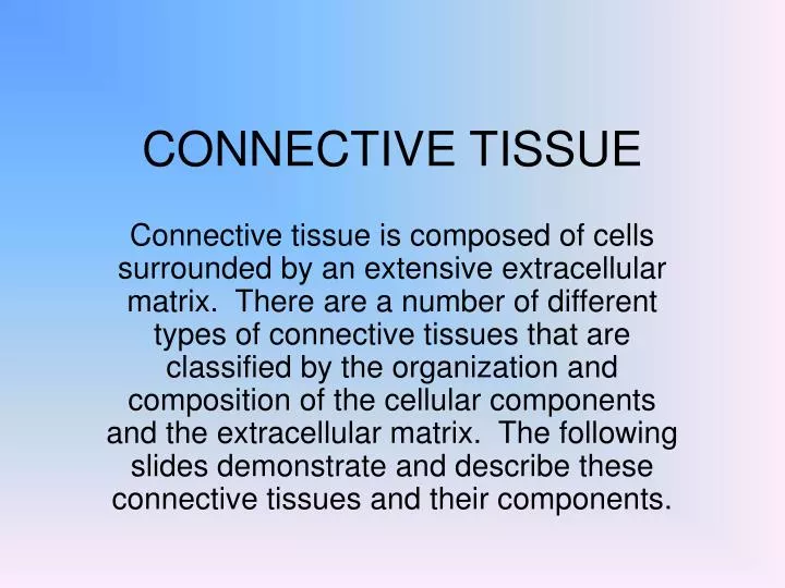
CONNECTIVE TISSUE Connective tissue is composed of cells surrounded by an extensive extracellular matrix. There are a number of different types of connective tissues that are classified by the organization and composition of the cellular components and the extracellular matrix. The following slides demonstrate and describe these connective tissues and their components.
Basic Connective Tissue Types • The basic connective tissue types include loose connective tissue, dense irregular connective tissue and dense regular connective tissue. These are classified based on the relative density and organization of the extracellular matrix components. There are a number of cell types that are resident to connective tissue. These are described in subsequent slides.
This shows a thin, plastic section of loose connective tissue from the oviduct stained with methylene blue- azure II. This type of connective tissue is very cellular and has few collagen fibers. The large elongated cell (arrow) is a fibroblast with an oval nucleus and much euchromatin.
This shows loose connective tissue from the mammary gland stained with hematoxylin and eosin. The loosely arranged collagen fibers (arrows) are acidophilic and lightly stained in this picture. A mast cell with a round nucleus is seen (MC) with a considerable amount of heterochromatin. Eosinophilic secretory granules fill the cytoplasm of this cell. MC
This shows loose fibrous connective tissue similar to that seen in the previous slide. Another mast cell (MC) is located near the center of the field. Thin, light-staining collagen fibers (arrows) are seen in this picture. MC
This shows a spread (not a tissue section) of a thin piece of mesentery illustrating a different view of loose connective tissue. The large, dark-staining cells are mast cells (MC) whose granules obscure the nuclei. The largest oval nuclei belong to mesothelial cells (Me), that form an epithelium on either side of the connective tissue. The smaller, oval nuclei belong to macrophages (Ma). These cells tend to have associated granular material in their cytoplasm. Other oval-shaped nuclei seen here are those of fibroblasts. Ma Me MC
This shows a transmission electron micrograph of a mast cell illustrating numerous dense secretory granules in the cytoplasm. Note also the elongated nucleus (Nu) with abundant heterochromatin. Nu
This shows another plastic section of loose connective tissue stained with methylene blue- azure II. A number of fibroblasts are seen (Fi) with oval euchromatic nuclei. A monocyte (Mo) is also seen with a darker, indented nucleus. A binucleate neutrophil (N) and a macrophage (Ma) with a small, light-staining nucleus are also seen. Mo N Fi
This shows two plasma cells (PC) located in the loose connective tissue of the mammary gland. These cells have round, eccentric nuclei with slightly basophilic cytoplasm (indicating the presence of RNP). The large, pale nucleus is that of a fibroblast while the darker nuclei are those of fibrocytes. PC
This shows adipose tissue stained with hematoxylin and eosin. The lipid in these fat cells is dissolved out during the fixation and embedding process. Only a thin rim of cytoplasm and the flattened nuclei (arrows) are seen.
This shows a developing adipose cell (arrow) stained with hematoxylin and eosin. Note the ring of cytoplasm and the rounded nucleus surrounding a lipid droplet. Acidophilic collagen fibers are seen around the cell.
This shows adipose cells preserved so that the lipid is retained in the cell and stained with osmic acid, hence the really dark appearance of the cells.
This shows loose connective tissue (CT) within a mucosal fold in the gall bladder. Numerous collagen fibers and abundant cells can be seen in the connective tissue. The cells cannot be readily identified at this magnification. (What type of epithelium is seen here?) CT
This shows a section of the spermatic cord illustrating dense irregular connective tissue. In dense connective tissues, the abundant collagen fibers form interwoven bands of tissue. In contrast to loose connective tissue, there are relatively few cells (mainly fibrocytes). On the edges of the tissue is a simple squamous epithelium (Ep). Ep
This shows dense irregular connective tissue at higher magnification than in the previous slide. The bundles of brownish collagen fibers can be seen coursing in different directions and different planes. A few thinner, darker staining elastic fibers can be seen in this section (arrows).
This shows dense elastic tissue of the aorta at low magnification. The thin elastic fibers (stained black) in the wall of the aorta (arrows) form layers separated by spaces. The collagen fibers are stained brown in this section.
This shows another example of dense irregular connective tissue from the urinary bladder at low magnification. This particular section has more cells than the previous example, but still fewer than the loose connective tissue seen earlier. Most of the nuclei belong to fibrocytes.
This shows another picture of dense irregular connective tissue from the urinary bladder. Note that the collagen bundles in the top of the field are more organized than those at the bottom.
This shows a section of the penis illustrating dense regular connective tissue stained with hematoxylin and eosin. Most of the collagen fibers (pink staining) are oriented parallel to each other. The nuclei of fibrocytes are present.
This shows dense regular connective tissue (CT) from a ganglion. On the left side of the field are elongated nuclei of nervous tissue (NT). The remainder of the field is occupied by dense collagenous fibers with few, flattened nuclei of fibrocytes. NT CT
This shows a transmission electron micrograph of a macrophage. The nucleus (Nu) is eccentrically located and is relatively heterochromatic. Note the lysosomes/residual bodies (R) in the cytoplasm as well as mitochondria, rough endoplasmic reticulum. Also, note the collagen fibrils outside the cell (C). Nu R C
This shows a transmission electron micrograph of a lymphocyte. Notice that the nucleus (Nu) occupies a large portion of the cell, is indented and has a fair amount of heterochromatin. The cytoplasm has only a few organelles and the plasma membrane tends to be irregular due to movement of the cell. Nu
CARTILAGE • Cartilage is a specialized form of connective tissue composed of cells called chondrocytes and their surrounding matrix. There are three types of cartilage that are distinguished based on their matrix characteristics.
This shows a light micrograph of hyaline cartilage located in the trachea. The pieces of cartilage (HC) are bordered by dense regular connective tissue of the perichondrium (P). The cartilage cells or chondrocytes (arrows) are located in lacunae (these are difficult to discern at this magnification). P HC
This shows a portion of the previous picture at higher magnification illustrating the chondrocytes in lacunae (arrows). The matrix surrounding the cells is composed of collagen fibers (too small too be seen here) and other components including glycosaminoglycans such as chondroitin sulfate.
This shows hyaline cartilage (HC) from the nasal cavity illustrating chondrocytes in their lacunae and the “glassy” appearance of the surrounding matrix (purple staining here). Note the organization of the dense regular connective tissue of the perichondrium (P) as compared to the irregular dense connective tissue of the surrounding connective tissue (*). HC P *
This shows a higher magnification of hyaline cartilage from the trachea. The chondrocytes on the right side of the field are not as mature and fill their lacunae (arrow) while those of the left have more space surrounding the cell.
This shows a section of elastic cartilage stained with a Verhoeff stain. Identify the perichondrium (P) surrounding the cartilage. Note the specifically stained elastic fibers (arrows) characteristic of this cartilage type. Also notice that the chondrocytes and their lacunae get larger as they progress from the edge of the cartilage towards its center. P
This shows a higher magnification view of elastic cartilage. The elastic fibers are more clearly seen in this picture as are the chondrocytes and their lacunae.
This shows elastic cartilage of the epiglottis stained with a Trichrome stain. Note that the collagen of the perichondrium (P) is stained green; however, the collagen type II within the cartilage does not stain. The elastic fibers are stained purple (arrows) P
This shows a light-stained section of fibrocartilage. The cartilage stains acidophilic due to the collagen fibers. Note that the chondrocytes and lacunae are small and often have some regular pattern to their organization (in rows). There is no perichondrium associated with this cartilage type.
This shows a higher magnification view of fibrocartilage illustrating the collagen fibers within the matrix surrounding the chondrocytes. This type of cartilage is often found between hyaline cartilage and dense connective tissue.
BONE • Bone is a specialized connective tissue with a mineralized extracellular matrix. Bone generally consists of three cell types including the osteoprogenitor cells, osteoblasts (osteocytes when differeniated) and osteoclasts. The following slides illustrate some of the features of bony tissue.
This shows a low magnification view of the end of a long bone. Above the epiphyseal plate (EP) is spongy bone. The outer portion of the shaft is compact bone (CB), which is laid down by appositional growth from the periosteum. The compact bone surrounds the spongy bone and bone marrow of the shaft. EP CB
This shows a cross-section of developing bone from a fetal finger. The outermost layer is the connective tissue of the periosteum (P). Small pieces of calcified cartilage are seen (blue staining) as are hypertrophying cartilage (arrows). Tendon P Spongy Bone Developing Compact Bone
This shows a higher magnification of the previous slide illustrating developing bone. Spongy Bone Calcified Cartilage
This shows red blood cells (RBC) in the bone marrow bordering pieces of calcified cartilage (arrows). The calcified cartilage (blue) is covered with osseous tissue (deep pink staining). RBC
This shows a scanning electron micrograph of bone surface. Note the Haversian Canals (HC), the concentric lamellae surrounding these and the lacunae of the osteocytes (arrows). HC
This shows a section of decalcified bone. The structural features are similar to that in the previous slide illustrating the Haversian system. Haversian Canal Osteocytes
This shows a transmission electron micrograph of an osteocyte surrounded by its lacuna. The processes extending from the cell body are located in canaliculi (arrows). The matrix of the bone surrounds the canaliculi.
This shows a section of decalcified bone and associated skeletal muscle (SM). The skeletal muscle is attached to the connective tissue of the periosteum (P). The large space is due to shrinkage of the periosteum during processing of the tissue. Small Haversian Canals can be seen within the bone (arrows). P SM
This shows a section of decalcified bone illustrating the connective tissue of the perioosteum (P). Osteoblasts are located along the inner edge of the periosteum (arrow). These cells will deposit and become entrapped in bony matrix (appositional growth). P Compact Bone
This shows another section of decalcified bone and associated skeletal muscle (SM). Note that the osteogenic layer of the periosteum is quite cellular (arrows) with basophilic osteoblasts located at the periphery of the bone. SM
This shows bony tissue (upper right corner) and the dense irregular connective tissue of the periosteum (P). Note that the indentation of the periosteum into the bone contains looser connective tissue. Lining the periosteum are basophilic-stained osteoblasts (Ob) Ob P
This figure shows decalcified bone (B) and dense connective tissue of an associated tendon (T) attaching to the periosteum. B T
This shows decalcified bone illustrating a resorption canal surrounded by the bony tissue. Osteoclasts (arrows) can be seen lying next to the bony tissue.
This illustrates bone marrow (BM) adjacent to inner circumferential lamellae of bone. The dark irregular areas (arrows) are the original calcified cartilage indicating that this was formed by endochondral ossification. BM
This illustrates bone marrow (BM) and the osteoblasts (Ob) lining bone. The osteoblasts extend into the beginning of a Volkmann’s Canal (VC), which carries blood vessels into the Haversian system. BM Ob VC
1 2 3 • This schematic illustrates the process of intramembranous ossification. Mesenchymal cells form an elaborate network (1). These cells then enlarge and differentiate into osteoblasts (2). The cells lay down an intercellular matrix an form bony spicules (3) covered by osteoblasts.
B • This is a decalcified section of the root of a tooth and its associated alveolar bone (B). The root of the tooth is covered by cementum (C) and collagen fibers which anchor the tooth into the bony tissue. C