Download
1 / 60
630 likes | 966 Vues
Digestive Systems. Campbell, 6 th edition, Chapter 41 Animal Nutrition. DIGESTIVE SYSTEMS. I. Digestion Food Types Feeding Mechanisms IV. Compartmentalization V. Stages of Food Processing VI. Types of Digestive Systems VII.Vertebrate Digestive System & Regulation. I . Digestion.

E N D
Digestive Systems • Campbell, 6th edition, Chapter 41 • Animal Nutrition
DIGESTIVE SYSTEMS I. Digestion • Food Types • Feeding Mechanisms IV. Compartmentalization V. Stages of Food Processing VI. Types of Digestive Systems VII.Vertebrate Digestive System & Regulation
I. Digestion • 1) fuel (chemical energy) • 2) raw organic materials (biosynthesis) • 3) essential nutrients • Homeostatic mechanisms manage these resources. • Know difference between undernourished and malnourished. Any nutritionally adequate diet satisfies three needs:
Essential Nutrients • 8 Amino Acids: tyrptophanmethionine valine threonine phenylalanineleucine isoleucinelysine (histidine is essential for infants) • Fatty acids: membrane structure unsaturated; linoleic acid • 13 Vitamins: coenzymes Fat-soluble: vitamins A, D, E, & K Water-soluble: B complex,Vitamin C • Minerals: building materials, cofactors Ca, P, S, K, Cl, Fe, Mg, Zn, I, Na,
I. Digestion • Chemical and mechanical breakdown of organic molecules into units small enough for the body to absorb. • These molecules provide: • 1) Energy resources • 2) Essential chemical elements • 3) Raw materials for anabolism
Food Types • Most animals are opportunistic feeders: 1) HERVIVORES – feed on autotrophs 2) CARNIVORES – eat other animals 3) OMNIVORES – both
III. Feeding Mechanisms Four major groups: • suspension-feeders Baleen whale, clam, oyster • substrate-feeders Leaf miner, earthworm • fluid-feeders Mosquito, leech • bulk-feeders Python, lion, bear
IV. Digestion is compartmentalized • Intracellular – digestive enzymes are secreted by cells & food is digested by enzymes within the cell • sponges (choanocytes) • Extracellular - digestive enzymes are secreted by cells into a digestive cavity • cnidarians (both intracellular & extracellular)
V. Stages of Food Processing • Ingestion • the act of eating or ingesting • Digestion • process of breaking food into small enough molecules for the body to absorb • Absorption • process of absorbing small molecules from the digestive compartment into bloodstream • Egestion • act of eliminating undigested materials from the digestive compartment
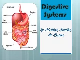
VI. Types of Digestive Systems • Channel network • Porifera • Incomplete (one-hole sac or gastrovascular cavity) • Cnidarians Platyhelminthes • Complete (two-hole sac or alimentary canal) • Nematoda Chordata
VI. Types of Digestive Systems • Incomplete Digestive Tract or gastrovascular cavity • functions in digestion (gastro) & distribution (vascular) • dual role: mouth = anus • Cnidaria Platyhelminthes
VI. Types of Digestive Systems • Complete Digestive Tract • Alimentary canal • food moves in one direction • tube is organized into specialized regions • Nematoda Chordata
Earthworm Digestive Tract
VII. Vertebrate Digestive System Oral cavity: mouth, tongue, teeth • digestion begins here • mastication= mechanical grinding action of teeth • food soften with saliva from salivary glands • bolus = moistened ball of food
Accessory Organs: Salivary Glands Saliva • contains mucus for lubrication and swallowing (1 - 1.5 L per day) • contains salivary amylase- hydrolysis of amylose • contains mucin, buffers, antibacterial agents • venom- secreted by the salivary glands of some vertebrates • Parotid, submaxillary, sublingual salivary glands
Pharynx (throat region) • swallowing is accomplished by the pharynx • an intersection leads to both esophagus and trachea; cartilage flap, epiglottis, covers the glottis & prevents choking (Figure 41.12) • passes bolus from mouth to esophagus • originates from a groove in the floor of the lungs • acts as a muscular pump in some worms (proboscis w/ pharynx)
From mouth to stomach: the swalling reflex and esophageal peristalsis
Esophagus • 25 cm long • muscularized passageway to stomach • peristalsis begins here rhythmic waves of contraction by smooth muscles in the wall of the canal • ruminants (cud-chewers), ruminating pouches, chambers of esophagus where fermentation occurs (cows produce 60 L saliva per day & burp 2 L gas/minute)
Layers of the Digestive Tube From esophagus to anus: • 1) mucosa – lines tube; glandular epithelium; villi; contains some smooth muscle; produces mucus • 2) submucosa – connective tissue, nerves, blood vessels, lymph • 3) muscularis externa – inner circular muscle & outer longitudinal muscle • 4) serosa – outer fibrous coating or visceral peritoneum
Cardiac sphincter • ringlike valve of smooth muscle that functions like a drawstring • controls entrance of food into stomach from esophagus
Stomach • collapsible muscular bag • suspended in abdominal cavity by folds of peritoneum called mesentery • functions in mechanical mixing of food with HCl and enzymes • fully distended, human stomach holds 2-4 liters of food
3 regions of the Stomach: • 1) cardiac – upper • 2) fundus – deep, storage • 3) pylorus – lower, empties into small intestine
Stomach • glucose and alcohol are absorbed in stomach • takes about 4 hours to empty stomach • chyme – semi-liquid mass; may back up in gastric pits and cause ulcers
Stomach • Rugae – folds of stomach with deep pockets, or gastric pits, contain • 1) mucous cells – secrete mucus for protection • 2) parietal cells – secrete HCl (pH 1.5-2.5) HCL kills most bacteria & living cells; erodes plant materials; initiates change of pepsinogen to pepsin • 3) chief cells – secrete pepsinogen (inactive) which is converted by HCL into active pepsin
Digestion in Stomach Digestion is regulated by hormones and the Autonomic Nervous System • Stomach hormone: Gastrin • produced in the presence of protein-containing food in the stomach • stimulates the release of gastric juices and muscular contractions of stomach & intestine • Blood sugar is regulated by pancreatic hormones insulin and glucagon
Digestion in Stomach Stimulation of epithelial cells of stomach mucosa increases secretion of gastric juice: • 1) mucous • 2) HCl • 3) pepsinogen • 4) renin(hydrolyzes milk) • 5) water
Pyloric sphincter • sphincter separating the stomach and small intestine • regulates the passage of material from stomach to small intestine
Small Intestine • digestion is completed here • most enzymatic hydrolysis and adsorption occurs here • surface area of small intestine is 300 m2;about the size of a double tennis court • mucosa has fingerlike projections, villi, which extend into the lumen • the villi have microvilli (cytoplasmic projections on the surface of epithelial cells) Figure 41.15
Small Intestine • Small intestine is 20-23’ long: • 1) duodenum 8-10” • 2) jejunum 8’ • 3) ileum 12’ • duodenum – most active in digestion • jejunum & ileum – absorption • study Figure 41.13: Enzymatic digestion
Digestion in Small Intestine Digestion is regulated by hormones and the Autonomic Nervous System • Duodenal hormones: • 1) secretin – stimulates pancreas & liver to secrete alkaline fluids • 2) cholecystokinin – triggers release of enzymes from pancreas and gall bladder (amylase, lipase, deoxyribonuclease, protease, etc.)
Activation of protein-digesting enzymes in the small intestine
Accessory Organs: Liver • largest internal organ = 3 lb chemical factory • processes food by the Hepatic Portal Vein delivered from digestive tract • variable nutrient levels in HPV while level in systemic circulation remains constant • though the liver performs many functions, cells of the liver function without division of labor