Download

1 / 15
150 likes | 259 Vues
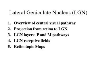
Anatomical Characteristics and Three-Dimensional Model of the Dog Dorsal Lateral Geniculate Body. By Inah Lee , Jejoong Kim & Choongkil Lee Department of Psychology Seoul National University. Introduction.

E N D
Anatomical Characteristics and Three-Dimensional Model of the Dog Dorsal Lateral Geniculate Body By Inah Lee , Jejoong Kim & Choongkil Lee Department of Psychology Seoul National University
Introduction In the domestic cat, the medial interlaminar nucleus (MIN), a medial subdivision of the lateral geniculate body (LGB), has a special role in dim-light vision. It almost exclusively represents a region of retina roughly coincident with the reflective tapetum (Lee C, Malpeli JG, Schwark HD, Weyand TG, J Neurophysiol 52,1984,p848- ), and cells of the MIN have relatively high luminance threshold at low adaptation levels (Lee D, Lee C, Malpeli JG, J Neurophysiol68,1992, p1235- ). The MIN has been described in many families of mammalian carnivores (Sanderson KJ, J Comp Neurol153,1974,p239- )suggesting that specialization for dim-light vision may be a general function of this structure. However, except for the cat, there is little specific information on functional organization and physiological characteristics of the MIN. As a preliminary step to investigate this relationship in another member of the carnivore family, we examined the laminar pattern of the entire LGB in one domestic dog breed, the Sapsaree. Anecdotal evidence suggests that the dog has excellent dim-light vision.
Our aim was to characterize anatomical features completely enough for guiding subsequent physiological studies. The method we used for determining laminar structure was to three-dimensionally reconstruct labeled retinal afferents following intraocular injection of an anatomical tracer, horseradish peroxidase (HRP). This strategy enabled us to unambiguously determine the subdivisions and layers of the LGB, to view complete three-dimensional features, and to estimate volume of each layer. Confirming previous results, the dog LGB consists of two subdivisions: the lateral geniculate nucleus (LGN) and the medial interlaminar nucleus (MIN). The MIN, however, was found to have more layers than previously thought: four orderly interdigitating layers receiving inputs from the contralateral and ipsilateral eyes.
Methods 1. Two male dogs, 6 and 12 months old, weighing 10 and 12 kg, were used. 2. The animals were tranquilized with ketamine (5 cc) and rompun (1 cc), 20 mg of HRP (Sigma) dissolved in 50 l of saline was injected into one eye. Animals survived 48 hrs after the tracer injection. All efforts were made to minimize animal suffering, to reduce the number of animals used. 3. Immediately after perfusion, a block containing the LGB (A4~A16.5) from both sides was removed from the brain. For alignment purposes, a needle hole and a knife cut groove shaped like a 'V'. 4. The block of the brain was frozen and cut into 17 um coronal sections with a sliding microtome. Every third section was processed for HRP (TMB method). Adjacent sections were counterstained with neutral red. The boundaries of LGN/MIN zones filled with HRP-labeled retinal afferents were traced under a microscope with the aid of a drawing tube, scanned into a computer, and then combined into 3-D, using a commercial software package (Voxwin 1.2.2, Voxar Co., U.K.).
5. Ambiguities in laminar assignment were resolved by the cytoarchitecture and by rotating three-dimensional views. 6. For comparison, laminar volumes of the cat were estimated using Figures of Sanderson(Sanderson KJ , J Comp Neurol 143, 1971,p101- ). His Figures were scanned into digital images as described above.
1. Composite photomicrographs of representative HRP-reacted coronal sections • Two ipsilateral MIN layers are clearly visible in this coronal section (left). We follow and extend the naming scheme that Guillery et al (Guillery RW, Geisert EE Jr, Polley EH, Mason CA , J Comp Neurol 194 ,1980,p117- )used for the cat; layers A, A1, C, C1, C2, and C3 for the LGN, anterior to posterior, and layers 1, 2, 3, and 4 for the MIN, medial to lateral. • A new finding of this study was that the dog MIN is organized into four orderly arranged layers: two contralateral (right) and two ipsilateral (left).
2. Laminar Pattern in Coronal Sections Upper: Representative sections of contralateral eye layers in the right MIN/LGN complex, posterior to anterior, selected from 79 slices. Each layer is color-coded according to the color-map in C. Five layers can be identified: layers A, C, C2 for the LGN, and layers 1, 3 for the MIN. Layer A is partially divided in posterior sections (a,b) into medial and lateral parts by a cleft (arrowheads in Figs. a,b). The lateral margin of layer A1, which defines the monocular segment, is roughly aligned with breaks in layers A and C (arrowheads in Figs. c,d and e) that presumably represent the optic disk. Lower: Representative sections of ipsilateral eye layers, posterior to anterior, selected among 68 slices. Sections made for the left side of the brain were flipped left to right to make the same orientation as in contralateral sections A. a-e; Four layers could be identified: A1, C1 for the LGN, and 2, 4 for the MIN.
3. A Summary of Laminar Structure Representative section showing the laminar structure of the dog LGB, combined from sections of a contralateral (Figure 2Ad) and ipsilateral (Figure 2Bd) layers. MIN consists of 4 orderly-alternating, contralateral and ipsilateral layers. The arrowhead points to the presumptive border of the monocular segment. Layer A is the largest layer of all. The MIN consists of four orderly-alternating layers of the contralateral and ipsilateral layers. The arrow points to a small laminar gap, beyond which layers A1 and C1 are no longer present and the number of layers abruptly changes from 5 to 3. This transition obviously marks the outer limits of binocular vision, and the gap probably corresponds to the optic disc representation13.
4. Sagittal and Horizontal Sections Representative parasagittal (upper) and horizontal (lower) sections of contralateral eye layers, lateral to medial. Ipsilateral eye layers are not shown. An arrowhead in a, points to a cleft which divides layer A into medial and lateral subdivisions. The projection column of the optic disc is oblique relative to the three cardinal planes, and it is not apparent in these sections. Since the break is oblique, it appears a line at the level of parasagittal section. However, it clearly is not the representation of the optic disk which lies at a more ventral level.
5. Three-dimensional Views of the LGN/MIN Complex Figure shows three-dimensional view of the computer-reconstructed LGB from sequentially-rotating perspectives. The combined complex appears like the letter ‘C’, with the convex part of the ‘C’ directed posteriorly.
6. Three-dimensional Views of the 4 MIN Layers MIN layer 1 contained the largest soma of all LGN/MIN layers, and a high density of labeled afferents from the contralateral eye. Layer 2 contained spindle-shaped large-sized somata and a high density of labeled afferents from the ipsilateral eye.
7. Volumetry (1 voxel = 0.0000504 mm3) Volumes of each LGN/MIN layer in mm3 . Volume percentage is given in parenthesis. Total volume of the LGB was 39.8 mm3. The LGN constituted 93 % and the MIN, 7 % of the whole complex. Contralateral layers occupied 78 %, and the ipsilateral, the remaining 22 %. The MIN constituted a similar fraction of the entire LGB on the two sides of the brain, 8 % on the ipsilateral side and 6 % on the contralateral side. Within the LGN, the ipsilateral projection was mostly directed to layer A1 (96%), with the remainder going to layer C1 (4 %). In comparison, layer A occupied 68 % of the total contralateral projection to the LGN.
9. Volume Comparison between the Dog and the Cat • Comparison of laminar volumes between the dog and the cat. Laminar volumes of the cat were estimated from figures of Sanderson (1971). Numbers are in mm3 and numbers in parentheses are volume percentage. Overall, volumes of the whole LGB are similar in the two species. • Compared with the cat, relative volume of the MIN, and ratio of layer A1 (ipsilateral) to layer A (contralateral) are lower in the dog.
Conclusions 1. Compared to the cat, the relative volume of ipsilateral layers was small. This perhaps is related to the extent of the partial decussation and direction of the optical axis; the dog has more lateral facing eyes, and thus a smaller binocular field compared to the cat. 2. The dog MIN consists of four orderly interdigitating layers; two contralateral layers (layers 1 and 3) and two ipsilateral layers (layers 2 and 4). General patterns of lamination in our breed (Sapsaree) were almost identical with those of unknown or mixed breeds used in previous studies (Rioch, 1929; Howard & Breazile, 1973; Morimoto, Kubota, Miyahara, & Kanaseki, 1984). However, we can not exclude the possibility that inter-breed differences in cytoarchitecture of the LGN/MIN exist, and that they account for the new MIN layers found in this study. 3. The quadrilaminar organization of the dog MIN raises several possibilities. First, layer 4 may represent the ipsilateral hemifield through the ipsilateral eye. Second, since MIN layers 3 and 4 layers are located near the border between the MIN and LGN, where the vertical meridian is mapped in the cat, they may represent a relatively more central region of contralateral visual space, and layers 1 and 2 a more peripheral zone via the two eye. Third, the two contralateral and two ipsilateral MIN layers may reflect the segregation of ON- and OFF-center cells, as has been shown in sublayers of the LGN in the mink (LeVay & McConnell, 1982), in the tree shrew (Conway, Schiller, & Mistler, 1980), and in the ferret (Stryker & Zahs, 1983). 4. The precise retinotopy of the dog LGN/MIN will need to be understood to allow a firm comparison of its role as a nocturnal specialization with that of the cat LGN/MIN.
Acknowledgement This research was supported by the Korea Ministry of Science and Technology. The dogs used in this study were kindly supplied by Dr. JiHong Ha at the Foundation for Sapsaree Conservation at Daegu, Korea. We thank Dr. Joseph Malpeli for constructive comments.