Download

1 / 37
390 likes | 652 Vues
Therapy for Challenging Wrist Conditions. Marianne Williams B.Sc.P.T., CHT. Objectives:. To discuss the therapy interventions for some wrist instability patterns. ANATOMY. Intrinsic wrist ligaments Volar Dorsal Extrinsic wrist ligaments Volar Dorsal. Intrinsic Wrist Ligaments-Volar.

E N D
Therapy for Challenging Wrist Conditions Marianne Williams B.Sc.P.T., CHT
Objectives: • To discuss the therapy interventions for some wrist instability patterns
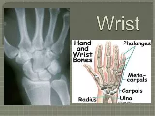
ANATOMY • Intrinsic wrist ligaments • Volar • Dorsal • Extrinsic wrist ligaments • Volar • Dorsal
Extrinsic Wrist Ligaments-Volar Capito-triquetral lig Radio-scapho--capitate lig
Extrinsic Wrist Ligaments-Dorsal Radio-triquetral lig
Classification • Carpal Instability Dissociative (CID) • A dissociation within the interosseous ligaments • Carpal Instability Non-dissociative (CIND) • Instability of a carpal row as a whole • Carpal Instability Complex (CIC) • A combination of the 2 dissociations • Carpal Injury Adaptive (CIA) • Secondary changes in the carpus which results from a non-union or malunion of the distal radius or carpal bones
CID - Pathomechanics • The proximal carpal row is the most common site • The scapho-lunate ligament disruption and subsequent scaphoid rotatory subluxation is the most common ligament injury pattern of the wrist • The clinical picture may range from complete dislocation to a subtle form of dynamic instability • The carpal mal-alignment results in increased stresses on the radio-scaphoid and capito-lunate joints
Clinical Evaluation • ROM • Grip Strength • Palpation over the Scapho-lunate(SL) ligament • Special tests :Watson’s test, SL Ballottement test • X-ray
Therapy Considerations • Kinematics: • As the wrist extends, there is a tendency for the scaphoid to supinate and the lunate to pronate, which effectively separates the palmar surfaces of the 2 bones. • The reverse occurs in palmar flexion • Berger 1996
Therapy Considerations (cont’d) • Kinetics: • The radial side of the wrist is responsible for the major load transfers across the wrist. In wrist neutral and neutral forearm rotation, 80% of the force is transmitted across the radiocarpal joint and 20% across the ulnocarpal joint with gripping. • Berger 1996
CID-Therapy Intervention • Comfortable AROM • No PROM into end range • No repetitive gripping • Isometric vs isotonic strengthening
Post-Operative Therapy • When ligaments are repaired, the patient may be immobilized for up to 8 weeks • Length of immobilization after a partial fusion is dependent upon radiographic healing • Initially AROM is started then PROM for extension sooner than flexion. PROM is for physiological range not accessory motion. • Therapy goals need to parallel the surgeon’s goals
Post-Operative Therapy (cont’d) • Functional wrist ROM is 40/40 for flexion and extension. • Wright et al, 1996 • Principles of strengthening are based on the kinetics. Initially start with isometrics and retraining the wrist extensors. Gradually progress to concentric strengthening. Grip strengthening is introduced judiciously.
Luno-triquetral Instability • Less common than the SL ligament dissociation • Alter the special tests in your clinical examination to this region • Similar approach by Surgeon and Therapist for both conservative and post-operative interventions as in SL dissociation
CIND: Clinical Characteristics • Volar sag on the ulnar side of the wrist • Painful clunk which occurs at the end range of ulnar deviation • Tenderness over the triquetral-hamate and capito-lunate intervals • Weakness of grip
Pathoanatomy • Ligamentous laxity or disruption results in the following: • Volar flexed orientation of the proximal carpal row in neutral deviation • Palmar translation of the distal carpal row • Ligaments involved: • Ulnar arm of the volar arcuate ligament • Dorsal radio-triquetral ligament
Pathomechanics • Loss of intimate midcarpal joint contact • Loss of the smooth transition of the proximal carpal row from a flexed to extended position as the wrist moves from radial to ulnar deviation • As the wrist moves from radial to ulnar deviation, the proximal carpal row stays flexed and the distal carpal row remains palmarly subluxed until the end range of ulnar deviation
Pathomechanics (cont’d) • A painful “clunk” occurs at the end range of ulnar deviation which represents abrupt snapping of the proximal carpal row into extension and reduction of the palmar translation of the distal carpal row. • Wright et al ,1984
Clinical Evaluation • Palpation over the triquetral-hamate and the capito-lunate intervals • Midcarpal shift test (Lichtman) • Passive translation of the midcarpal joint while stabilizing the distal forearm and applying a palmar or dorsal directed force to the carpus
CIND-Conservative Management • This is attempted initially • Immobilize – cast for 6 weeks • Then wrist cock-up splint and initiation of AROM • PROM OFTEN NOT NECCESSARY
Midcarpal Stabilization Splint • Dorsally directed pressure on the pisiform reduces the ulnar sag and corrects the volar flexed position of the proximal carpal row • Midcarpal dynamics are corrected and wrist clunk is eliminated
Midcarpal Stabilization Splint • Splint allows nearly full extension, radial and ulnar deviation and limits flexion
Exercise Approach • Midcarpal joint pathomechanics might also be corrected by dynamic muscle action • Dynamic muscle compression achieved by activation of the flexor carpi ulnaris (FCU) and the hypothenar muscles reproduces the normal joint contact forces in the absence of adequate ligament support
Abd DM FCU ECU
Abd DM ECU FCU
Exercise Approach (cont’d) • Strengthening of both FCU and ECU, would seem to be important in counteracting the volar sag that occurs prior to moving into ulnar deviation • Position of the forearm – supination • Isometric vs Isotonic exercises
Case Presentation • 22 year old right-handed cashier with idiopathic right dorsal wrist pain • Diagnosis: Midcarpal Instability • Right wrist was immobilized in a cast x6/52 • April 22, 2003 Right wrist cock-up splint fabricated and patient initiated AROM exercises • Sept 17,2003 midcarpal stabilization splint made for the right wrist
PRWE Results • Sept. 17,2003 Pain: 37/50 Function: 29.5/50 Total: 66.5/100 • Right midcarpal stabilization splint made • Nov. 4, 2003 Pain: 22/50 Function: 11.5/50 Total: 33.5/100
Surgical Intervention • At our centre the approach is dorsal • The Hand Therapist should be cognizant that if a procedure is designed to stabilize tissue and the prolonged post-operative immobilization is to limit motion, then therapy principles should parallel these goals.
CIC-Perilunate Dislocation • Surgery is the most successful form of medical management • Post-operative therapy is similar to that of SL or LT repair • However, ligament repairs are more extensive and more dissection required • Therefore, long extensor adhesions are more common • Significant wrist motion is lost post-operatively
CIA-Carpal Injury Adaptive • Secondary changes in the carpus have developed from a non-union or malunion of the distla radius or carpal bone • The pattern is treated by correcting the deformity eg Distal radius corrective osteotomy • This post-operative therapy focuses on early AROM
Conclusion “A good axiom to keep in mind is that a painless, stable wrist with functional ROM is superior to one with full ROM that is unstable and painful” Hendrycks et al, 2001