Download
1 / 40
400 likes | 682 Vues
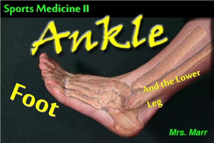
Sports Medicine II. And the Lower Leg. Mrs. Marr. Foot. TIBIA. FIBULA. TALUS. CALCANEUS. THE 4 BONES OF THE ANKLE JOINT. ANKLE LIGAMENTS – MEDIAL. Deltoid Ligament Complex 4 ligaments Broad Flat Overlapping = STRONG!. D. A. C. B. ANKLE LIGAMENTS - LATERAL.

E N D
Sports Medicine II And the Lower Leg Mrs. Marr Foot
TIBIA FIBULA TALUS CALCANEUS THE 4 BONES OF THE ANKLE JOINT
ANKLE LIGAMENTS – MEDIAL • Deltoid Ligament Complex • 4 ligaments • Broad • Flat • Overlapping • = STRONG! D A C B
ANKLE LIGAMENTS - LATERAL • Lateral Collateral Ligaments • ATF • Anterior • From Talus to Fibula • Weakest of 3 ligaments • PTF • Posterior • From Talus to Fibula • Strongest/Deepest of 3 ligaments • CF • Anterior • From Calcaneus to Fibula • Largest; Strong and Cord-like
ANKLE MUSCLES: ANTERIOR SIDE • TIBIALIS ANTERIOR • Muscle starts @ top of Tibia • Tendon crosses over Ankle Joint @ Talus • Attaches at the base of the 1st foot bone • Cross over at joint allows for multiple motions • Major Motion: • Dorsiflexion of the ankle joint • Inversion of the foot • Prevents the forefoot slapping AND scrapping the ground
ANKLE MUSCLES: LATERAL SIDE • PERONEAL GROUP • 3 muscles (peroneus brevis/longus/tertius) • Muscle group starts @ top of Fibula • Peroneal tendon hooks around the back of Lateral Malleolus • Insertion of Peroneal Tendon is at the base of the 5th foot bone • Major Motion: Eversion of the Foot @ the Ankle
ANKLE MUSCLES: POSTERIOR • GASTROCNEMIUS • Muscle starts on distal femur • 1 muscle with two points of origin • Achilles Tendon is other attachment • Crosses two joints Major Motion: Plantarflexion @ the Ankle • ACHILLES TENDON • Large Tendon/Cord from Gastroc. • Inserts firmly at Calcaneus • Largest, Strongest Tendon in Body • Combination of Gastroc and Soleus Tendons
Compartments of the Leg • Anterior • Lateral (peroneal) • Deep posterior • Superficial posterior
Anterior Compartment Musculature • Tibialis anterior • Extensor digitorum longus • Extensor hallucis longus • Peroneus tertius
Tibialis Anterior • DF and inversion • O: lateral tibial condyle and shaft • I: medial/plantar 1st cuneiform and metatarsal • N: deep peroneal
Extensor Digitorum Longus • Extension of 2nd-5th MP joints, assists with eversion and DF • O: lateral tibial condyle, proximal ¾ of anterior fibula • I: via 4 tendons into distal phalanges of 2nd-5th toes • N: deep peroneal
Extensor Hallucis Longus • Extension of 1st MP and IP joints • O: middle 2/3 of anterior fibula • I: base of distal 1st phalanx • N: deep peroneal
Peroneus Tertius • Eversion of foot, assists in PF • O: distal 1/3 of anterior fibula • I: dorsal base of 5th metatarsal • N: deep peroneal
Lateral Compartment Musculature • Peroneus longus • Peroneus brevis
Peroneus Longus • Eversion of the foot, assists with PF • O: lateral tibial condyle, fibular head, upper 2/3 of lateral fibula • I: lateral base of 1st metatarsal, lateral and dorsal aspect of 1st cuneiform • N: superficial peroneal
Peroneus Brevis • Eversion of the foot, assists with PF • O: distal 2/3 of lateral fibula • I: styloid process at base of 5th metatarsal • N: superficial peroneal
Superficial Posterior Compartment Muscles • Gastrocnemius • Soleus • Plantaris
Gastrocnemius • Ankle PF, assists knee flexion • O: medial head – posterior medial femoral condyle, lateral head – posterior lateral femoral condyle • I: calcaneus via Achilles tendon • N: tibial
Soleus • Ankle PF • O: posterior fibular head, upper 1/3 of posterior fibular, soleal line on posterior tibial shaft, middle 1/3 of medial tibial border • I: calcaneus via Achilles tendon • N: tibial
Plantaris • Ankle PF, assists knee flexion • O: distal supracondylar line of lateral femoral condyle, femoral popliteal surface • I: calcaneus via Achilles tendon • N: tibial
Deep Posterior Compartment Musculature • Flexor Hallucis Longus • Flexor Digitorum Longus • “Tom, Dick, AND Harry” • Tibialis posterior
Tibialis Posterior • Inversion of the foot, assists with PF • O: posterior/lateral tibia, upper 2/3 of medial fibula • I: navicular tuberosity, via slips into sustentaculum tali, cuneiforms, cuboid and bases of 2nd-4th metatarsals • N: tibial
Flexor Digitorum Longus • Flexion of 2nd-5th PIP/DIP/MP joints, assists with foot inversion and PF • O: posterior medial 2/3 of distal tibia • I: plantar surface of base of 2nd-5th distal phalanges • N: tibial
Flexor Hallucis Longus • Flexion of 1st IP joint, assists with flexion of 1st MP joint, foot inversion and PF • O: posterior/distal 2/3 of fibula • I: plantar surface of 1st proximal phalanx • N: tibial
Syndesmosis Interosseous membrane (Syndesmosis) isn't a compartment but ligamentous sheathe that holds the tibia and the fibula together.
Nerves and Blood Vessels • Nerves: • Peroneal N. • Tibialis Anterior/ Posterior N. • Saphenous N. • Blood Vessels • Dorsal Pedal A. • Posterior Tibial A. • Greater/ Lesser Saphenous V.
Neuroanatomy • Anterior compartment • Deep branch of Peroneal nerve • Lateral compartment • Superficial branch of Peroneal nerve • Deep posterior compartment • Tibial nerve • Superficial posterior compartment • Tibial nerve
Deep Branch of Peroneal Nerve • Branches from common Peroneal nerve near fibular head • “Dives” into anterior compartment
Superficial Branch of Peroneal Nerve • Branches from common Peroneal nerve near fibular head • Stays superficial and lateral in lateral compartment
Tibial Nerve • Runs in fascial sheath between deep and superficial posterior compartments • Provides innervation to both, but not “in” either
Vascular Anatomy • Anterior compartment • Anterior tibial artery • Lateral compartment • Peroneal artery • Deep posterior compartment • Posterior tibial artery • Superficial posterior compartment • Posterior tibial artery
Anterior Tibial Artery • Traverses similar path to deep Peroneal nerve • Terminating as dorsal pedal artery
Peroneal Artery • Branches off of posterior tibial artery
Posterior Tibial Artery • Runs in fascial sheath between deep and superficial posterior compartments • Provides vascular supply to both, but not “in” either
Special TestsThe Squeeze Test • Squeeze test • check malleolus • Check tibia and fibula • May indicate FX • Feel for any abnormalities • Feeling for grinding or movement
Special TestsAnterior Drawer/Tilt Anterior drawer tests should always be performed with the knee bent to eliminate the Achilles and Gastrocnemius muscles from providing any stability to the ankle. A lateral talar tilt test can be conducted at the same time.
Special TestsFunctional Tests • Functional tests (Return to play) a) walking - check gait b) toe raises 1) both feet 2) one foot c) jump and land on both feet and then on one foot