Download
1 / 11
110 likes | 340 Vues
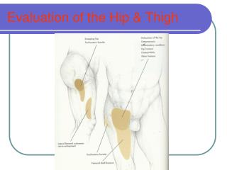
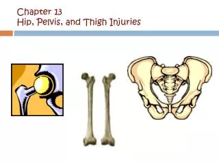
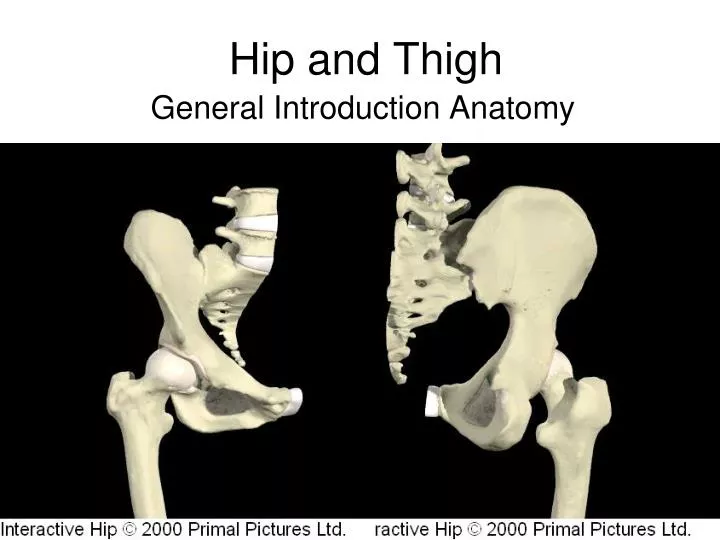
Hip and Thigh. General Introduction Anatomy. Hip Joint. Ball and Socket Ball = Femoral Head Socket = Formed by the three Pelvic Bones Socket called the Accetabulum Much more stable than the shoulder ball and socket. Hip Joint Stability. Deep Socket Femoral Head fits inside

E N D
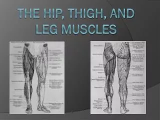
Hip and Thigh General Introduction Anatomy
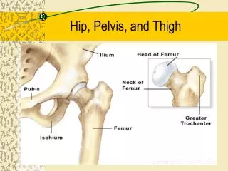
Hip Joint • Ball and Socket • Ball = Femoral Head • Socket = Formed by the three Pelvic Bones • Socket called the Accetabulum • Much more stable than the shoulder ball and socket
Hip Joint Stability • Deep Socket • Femoral Head fits inside • Deepened & reinforced by a fibrocartilagionous rim • Labrum Acetabulare • Encased by a fibrous synovial capsule • Three Strong ligaments support the joint • Iliofemoral & Pubofemoral – Anteriorly • Ischiofemoral - Posteriorly
Hip Bursae • Many bursae surround the hip • Two are very important and often injured/damaged in athletics • Trochanteric Bursa • Posterior to the Greater Trochanter • Deep to the Gluteus Maximus & Tensor fascia lata muscles • Iliopsoas Bursa • Between the joint capsule and Iliopsoas anteriorly
Iliopsoas, Sartorius, Rectus Femoris, Adductors Gluteus Maximus, Hamstrings Adductors, Medial Hamstrings Tensor fascia lata, Gluteus Medius & Minimus Tensor fascia lata, Gluteus Medius & Minimus, Adductors, Iliopsoas Gluteus Maximus, Piriformis, Obturartor, Gemellus Primary Flexors Primary Extensors Primary Adductors Primary Abductors Internal Rotators External Rotators Hip Muscles
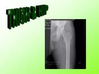
Femur • Largest, Longest & Strongest bone • Shaft slightly bowed outward & forward • Accommodate stress of locomotion & weight bearing • Femoral Neck • Narrows from the Femoral Head • Just lateral and angled down of the Femoral Head • Weakest point • 2 Large prominences on the superior shaft • Greater Trochanter • Lesser Trochanter • Attachment points for ligaments & Muscles
Pelvis • Pelvic Ring / Girdle • 7 bones • Sacrum • Posterior • Flattened fused spinal column vertebrae • Base support of the verteral column • Coccyx • 3-5 fused vertebrae • Terminal portion of the spinal column
2 Innominate bones • Sides of the pelivs • 3 fused bones • Amphiarthrosis joints • Curve to form bowl • Ilium • Ischium • Pubis
Pubic Symphysis Anterior joint Joins the two innominate pelvic wings Iliac Crest Outer uppermost ilium Margin 4 Iliac Spines Muscular Attachments ASIS, AIIS PSIS, PIIS Ischial Tuberosity Hamstring attachment Weight Bearing structure for sitting Pubic Tubercle Adductor Attachment Pelvic Landmarks