Download
1 / 104
1.06k likes | 1.61k Vues
The Client with Alterations in Cardiac Output . Lecture 9/26/05 Sherry Burrell, RN, MSN Rutgers University Nursing III. Assessment Parameters. Cardiac Output Measures the effectiveness of the heart’s pumping abilities.

E N D
The Client with Alterations in Cardiac Output Lecture 9/26/05 Sherry Burrell, RN, MSN Rutgers University Nursing III
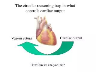
Assessment Parameters • Cardiac Output • Measures the effectiveness of the heart’s pumping abilities. • CO is defined as the amount of blood that leaves the heart in one minute. CO = Stroke Volume (SV) X Heart Rate (HR) • Normal CO: Approximately 4-8 liters/minute • Cardiac Index: CO per square meter of BSA • CO ÷ body surface area = CI
C0= SV x HR • Stroke Volume (SV) • The amount of blood that leaves the heart with each beat or ventricular contraction. • Not all blood ejected • Normal Adult 70 ml / beat • Ejection Fraction (EF) • The percentage of end-diastole blood actually ejected with each beat or ventricular contraction. • Normal adult 55-70% (healthy heart)
Stroke Volume • Three factors regulate stroke volume: • Preload • The degree of stretch of the ventricle at the end of diastole. • Contractility • Force of ventricular contraction (systole); inotropy. • Afterload • The amount of resistance the ventricular wall must overcome to eject blood during systole.
Stroke Volume Cont., • Preload • The degree of ventricular stretch at end-diastole • The Frank-Starling Law of the Heart • Preload = Contractility (to a point) • Factors Affecting Preload • Circulating volume • Body positioning • Atrial systole or “kick” • Medications • Diuretics (i.e. Lasix) • ACE Inhibitors (i.e. Vasotec) • I.V. Fluids Starling Curve
Stroke Volume Cont., • Contractility • Positive inotropic agents • Force of contraction • Negative inotropic agents • Force of contraction • Factors that affect contractility • Autonomic nervous system (ANS) • Medications: • Digoxin (Lanoxin) • Beta-adrenergic blockers (i.e. metoprolol ) • Calcium channel blockers (i.e. verapamil )
Stroke Volume Cont., • Afterload • Resistance to ventricular ejection during systole • Factors that affect afterload • Outflow impedance • Left side • High systemic blood pressures (SVR) • Aortic valve stenosis • Right side • High pulmonary blood pressures (PVR) • Pulmonary valve stenosis • Diameter of arterial vessels • Blood characteristics • Medications: • ACE (angiotension converting enzyme) inhibitors
CO = SV x HR • Heart Rate: beats per minute (bpm) • HR = CO (to a point) • HR >160 bpm = CO • Leads to inadequate diastolic filling time = time for coronary artery filling and an increase workload of the heart. • Factors that affect heart rate • ANS • Medications • Atropine sulfate • Digoxin (Lanoxin) • Beta-adrenergic blockers / calcium channel blockers
Assessment Considerations • General Cardiac Symptoms • Fatigue • Chest pain or discomfort • Palpitations • Shortness of breath • Edema • Weight gain • Dizziness • Syncope, loss of consciousness
Assessment: Special Populations • Gerontologic Considerations • Heart function is adequate at rest; limited ability to respond to stress and takes longer to return to baseline. • Decrease sensation of chest pain; tend to be under quantified or even absent. • Gender Considerations • Women: • Smaller hearts and coronary arteries • Tend to present with “atypical symptom” of CAD • Other Considerations • Diabetes mellitus and cardiovascular disease • Increased threat; decreased symptoms !!
Laboratory Analysis • Serum Enzymes • Blood Chemistry • Lipid Studies • Electrolytes • Renal Function Studies • Coagulation Studies • Hematologic Studies
Creatine Phosphokinase (Total CK / CPK) Non-Specific: enzyme elevated with damage to heart or skeletal muscles and brain tissue. Elevates in 4 to 8 hours Peaks in 15 to 24 hours Returns to normal in 3 to 4 days Creatine Phosphokinase Isoenzyme(CPK-MB) Specific: isoenzyme of CPK; elevated with cardiac muscle damage. Elevates in 4 to 8 hours Peaks in 15 to 24 hours Returns to normal in 3 to 4 days Serum Enzymes: Cardiac
Cardiac Enzymes • Myoglobin • Non-specific: a heme protein found in muscle tissue; elevated with damage to skeletal or cardiac muscle. • Elevates in 2 to 3 hours • Peaks 6-9 hours • Returns to normal 12 hours • Lactic Acid Dehydrogenase (LDH) • Non-specific: enzyme elevated with damage to many body tissues. (i.e. heart, liver, skeletal muscle, brain and RBC’s); Not frequently used today. • Elevates in 1 to 3 days • Peaks in 2 to 5 days • Returns to normal 10 to 14 days
Cardiac Enzymes Cont., • Troponin I / T • Specific: a contractile protein released with cardiac muscle damage; not normally present in serum. • Elevates in 4 to 6 hours • Peaks in 10 to 24 hours • Returns to normal in 10 to 15 days • Sensitivity superior to CK-MB within the first 6 hours of event. • Has replaced LDH for client’s who delay seeking treatment.
Other Serum Enzymes • C-Reactive Protein • Protein marker of acute inflammatory reactions • Increased serum levels associated with increased risk of acute cardiovascular events. • Homocysteine • Amino acid; presence in serum suggests increased risk of cardio-vascular events. • Natriuretic Peptides • Hormone-like substances released into bloodstream with cardiac chamber distention. • Atrial Natriuretic Peptide (ANP) • Brain or B-type Natriuretic Peptide (BNP)
Blood Chemistry Analysis • Lipoprotein (Lipid) Profile • Total Cholesterol • Normal < 200mg/dl • Triglyceride • Normal < 150 mg/dl • Low Density Lipoproteins (LDL) • Normal <130 mg/dl / “Optimal” <100mg/dl • High Density Lipoproteins (HDL) • Normal: > 40 mg/dl > 60 mg/dl cardio-protective
Blood Chemistry Analysis Cont., • Serum Electrolytes • i.e. Na, K, Ca and Mg • Glucose / Hemoglobin A1C • Coagulation Studies • PTT / aPTT • PT / INR • Hematologic Studies • CBC • Renal Function Studies • BUN • Creatinine
Diagnostic Testing • Electrocardiography * • 12-Lead EKG • Continuous bedside monitoring • Ambulatory monitoring • Stress Tests • Thallium Scans • Echocardiograms • Cardiac Catheterizations * Previously Discussed
Cardiac Stress Tests • Stressing the heart to monitor performance • Assists in Determining • Coronary artery disease • Cause of chest pain • Functional capacity of heart • Identify dysrhythmias • Effectiveness of medications • Establish goals for a physical fitness routine
Cardiac Stress Tests Cont., • Types of Stress Tests • Exercise • Treadmill (most common) • Bike • Arm crank • Pharmacological • Vasodilating agents to mimic the effects of exercise • Persantin • Adenosine • Mental / Emotional (new; under investigation) • Simulated public speaking • Mental arithmetic test
Cardiac Stress Tests Cont., • Thallium Scan • Often combined with stress tests • Radiological exam to assess how well the coronary arteries perfuse the myocardium. • Images are taken 1 to 2 minutes prior to end of stress test and again 3 hours later. • Nursing Considerations • NPO • IV Access
Cardiac Stress Tests Cont., • Nursing Considerations • Explain procedure to client • Maintain NPO status 4 hour before test • Instruct client to avoid stimulants (i.e. chocolate, caffeine and cigarettes) • Hold certain medications before testing • Exercise: i.e. beta-adrenergic blockers • Pharmacologic: i.e. Theophylline (24-48 hours prior) • I.V. access must be obtained
Echocardiogram • Ultrasound procedure of the heart combined with an electrocardiogram (EKG). • Assesses • Cardiac geometry (size & shape) • Motion of structures (chamber walls / valves) • Simultaneous EKG assists in interpretation • Can be done in conjunction with stress testing • Referred to as a stress echocardiography or exercise echocardiography
Echocardiogram Cont., • Types of Echocardiograms • Transcutanoeous • Non-invasive / painless • Transesphogeal (TEE) • Invasive / Clearer images • Nursing Considerations: • Explain procedure • NPO 6 hours prior procedure • I.V. access • NPO 4 hours post-procedure • Monitor for complications
Cardiac Catheterization • “Gold Standard” of cardiac diagnostics • Invasive procedure to assess • Cardiac chamber pressures & oxygen saturations • Detect congenital or acquired structural defects • Ejection fraction • Often Includes: • Coronary arteriography: to assess coronary artery patency • Using X-ray technique called fluoroscopy • Requiring the use of I.V. contrast / dye
Cardiac Catheterization Cont., • Nursing Care • Prior to procedure • Explain procedure • NPO prior to procedure (8 to 12 hours) • Check allergies (I.V. dye / shellfish / iodine) • Laboratory tests • During procedure • I.V. access • Hemodynamic monitoring • Arterial and venous access via catheters (sheaths) • Femoral (most common) or brachial
Cardiac Catheterization Cont., • Post-Procedure Nursing Care • Maintain Client Bedrest for 6 to 8 hours • Extremity straight & HOB up < 30 degrees • Maintain Adequate Hydration • IV Fluids (if ordered) • Encourage Fluids • Frequent Monitoring For Complications • Vital signs • Puncture site • Distal pulses • Laboratory results
The Client with Alterations in Cardiac Output Lecture II 9/30/05 Sherry Burrell, RN, MSN Rutgers University Nursing III
Coronary Artery Disease (CAD) • An insidious, progressive disease resulting in coronary artery narrowing or total occlusion. • Atherosclerosis • Most common cause of CAD • The abnormal accumulation of plaques on the vessel wall; involves inflammatory process. • Causes narrowing then eventually blockages in the coronary arteries that reduces myocardial blood flow = CAD • Asymptomatic until 75% occlusion of coronary artery lumen.
Coronary Artery Disease Cont., • Basis of CAD Management • Framingham Study (1948- cont. today) • Identified specific risk factors and life-style habits that increase one’s risk for developing atherosclerotic heart disease.
Modifiable Cigarette smoking Hypertension (HTN) Hyperlipidema Physical inactivity Diabetes Mellitus Obesity Stress / Anxiety Diet Non-Modifiable Increasing Age Males >45 years old Females >55 years old Gender Affects both men and women; #1 killer is U.S. Genetics Strong genetic component Ethnicity Non-whites increased incidences versus whites CAD: Risk Factors
Smoking Cessation Diet Exercise Weight Management Cholesterol Management Lipid Profile Normal: every five years Medications: i.e Zocor, Crestor & Niaspan HTN Management BP Screenings Medications: i.e. antihypertensives & diuretics. Diabetes Management Blood glucose testing Medications: i.e. oral hypoglycemics & insulin CAD: Interventions
Angina Pectoris • As CAD progresses the atherosclerotic plagues become significant, reducing blood flow to portions of the myocardium = Ischemia. • Myocardial ischemia clinically manifests most often as angina or chest pain. • Angina pectoris is defined as myocardial ischemia without cellular death. • Imbalance between myocardial oxygen supply and demand
Myocardial Oxygen Supply and Demand Balance Demand Supply O2 O2 Preload Afterload Arterial Oxygen Content Coronary Artery Blood flow Heart Rate Contractility
Precipitating Factors: Angina • Any situation where oxygen demands are increased: • Physical exertion • Tachycardia • Dysrhythmias • Cold weather • Eating a heavy meal • Stress or emotional states
Angina Pectoris • Signs and Symptoms • Chest Discomfort or Pain • Can occur anywhere in chest; most commonly behind sternum; poor localization • Pain may radiate to the back, arms (left most common), shoulder, neck or jaw. • Described as pressure, tightness or burning sensation • Often precipitated by physical exertion or stress • Maybe associated with a few of the following symptoms: • SOB, weakness, anxiety, diaphoresis, N/V, dizziness or numbness in upper extremities
Types of Angina • Stable Angina • Predictable, consistent pain with physical exertion & relieved with rest; “my usual chest pain” • Rest & NTG; can be managed medically for years • Unstable Angina • Last longer than stable angina, new onset or increased frequency / intensity of symptoms; pain at rest • Preinfarction or Crescendo Angina • Lasting longer than 15 minutes /unrelieved by NTG x3 is a medical emergency! • Call 911 / hospitalization for management
Types of Angina Cont., • Variant / Prinzmetal Angina • Pain at rest; maybe cyclic, + ST segment elevation (reversible); usual cause is coronary artery vasospasm with or without atherosclerotic plaques • Nitrates & calcium channel blockers • Silent Angina • No signs or symptoms; + ST segment elevation • Nitrates, beta blockers, calcium channel blockers & lifestyle changes
Management: Unstable Angina • Goals of Medical Management • Increase O2 supply & decrease O2 demand to the myocardium. • Prevent MI and death • To actively intervene !! • 12-Lead Electrocardiogram (EKG) • Significant CP without EKG changes; + changes treated as an MI. • Laboratory Tests • Electrolytes • Cardiac Enzyme Panel • Rule-out MI: every 8 hours x 3 / 6 hours x4
Management: Unstable Angina • Relief of Chest Pain: “MONA” • Morphine (drug of choice) • Oxygen • Nitroglycerine • Increase Coronary Artery Blood Flow • Antiplatelet medications • ASA • Glycoprotein (GP) IIb/IIIa Inhibitors • Heparin • Percutaneous Coronary Intervention (PCI)
Management of Unstable Angina Demand Supply O2 Contractility • HR • Afterload Preload O2 NTG ACE I Morphine Beta Blockers Ca Channel Blockers ACE I Blood Flow Open Occluded Arteries NTG Ca Channel Blockers ASA Anticoagulants Morphine PCI
Nursing Interventions: Unstable Angina • Early Identification of Chest Pain • Assessment of Chest Pain • Chest Pain: Intensity- Scales (0-10) & Characteristics- “OLD CART” • Mentation, overall tissue perfusion • Vital signs, heart rhythm, pulse oximetery • Diagnostics: 12- lead EKG and Laboratory tests • Management of Chest Pain =“MONA”
Nursing Interventions: Unstable Angina • Calm Environment • Anxiety and fear of impending doom (death) • Activity Restrictions • Avoid the valsalva maneuver • Patient Education • Risk factors for CAD • Signs and symptoms of angina • Medications • When to call the doctor • Stress management techniques See pp.403 box 16-8 Thalen
Acute Coronary Syndromes (ACS) • Umbrella term to describe a wide range of clinical presentations of CAD from unstable angina to acute myocardial infarction (MI). • Continuum, Not separate disorders! Acute Myocardial Infarction Unstable Angina
Myocardial Infarction (MI) • An MI is defined as irreversible necrosis (death) of myocardial tissue, resulting from an abrupt decrease or total lack of coronary blood supply. • An abrupt and severe disruption of O2 supply and demand to the myocardium. • Causes: • Coronary artery thrombosis (most common) • Coronary artery vasospasm • Cocaine • Trauma • Severe and abrupt hypotension
Myocardial Infarction (MI) Cont., • Signs and Symptoms: • Chest Pain • Severe and unrelenting substernal chest pain; often radiating to the back, left arm or jaw. • Lasting for 30 minutes or more • Only relieved by opioids • Occurs without a know precipitating event; usually occurring in the morning • Associated Symptoms • SOB, weakness, anxiety, diaphoresis, N/V, dizziness or numbness in upper extremities.
Myocardial Infarction Cont., • Pathophysiology • Irreversible cell death within 20-40 minutes of cessation of blood flow. • Wavefront of cellular death proceeds from endocardium to epicardium. • EKG changes associated with an MI: • Ischemia: T wave inversion • Injury: ST segment elevation • Infarction: Pathological Q waves
Types of Myocardial Infarctions • Classified according to muscle layer affected: • Q wave MI • Transmural: full thickness muscle wall necrosis • Often associated with a more prolonged MI • Non-Q wave MI • Partial-thickness muscle wall necrosis • Often associated with smaller, less complete occlusions. • i.e. Subendocardial- necrosis of the inner 1/3 to 1/2 of the muscle wall.
Types of Myocardial Infarctions • According to anatomical location • Left Ventricle • Anterior Wall • Left Anterior Descending (LAD) • Associated with left ventricular failure, pulmonary edema & cardiogenic shock • Inferior Wall • Right Coronary Artery (RCA) • Associated with dysrhythmias & conduction disturbances • Posterior Wall • RCA or Circumflex Artery • Right Ventricle • Portion of the RCA; Rare
Complications: Post-Acute MI • Dysrhythmias (Most Common) • Sinus Bradycardia • Occurs in about 40% of clients after an acute MI • Sinus Tachycardia • Must be corrected !! • Atrial • PAC’s or Atrial fibrillation common • Ventricular • PVC’s and ventricular tachycardia (VT) • AV Heart Blocks • Most common with inferior wall MI