Download
1 / 9
320 likes | 2.16k Vues
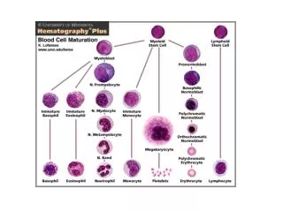
GRANULOPOIESIS AND THE FUNCTIONS OF GRANULOCYTE AND MONOCYTE. Dr. M.A. NDAKOTSU DEPARTMENT OF PATHOLOGY (HAEMATOLOGY). GRANULOPOIESIS. Pluripotent stem cell CFU-GM CFU-G CFU-M Myeloblast Monoblast Promyelocyte Promonocyte Myelocyte Monocyte Metamyelocyte

E N D
GRANULOPOIESIS AND THE FUNCTIONS OF GRANULOCYTE AND MONOCYTE Dr. M.A. NDAKOTSU DEPARTMENT OF PATHOLOGY (HAEMATOLOGY)
GRANULOPOIESIS Pluripotent stem cell CFU-GM CFU-G CFU-M Myeloblast Monoblast Promyelocyte Promonocyte Myelocyte Monocyte Metamyelocyte Band neutrophil Neutrophil
CONTROL OF GRANULOPOIESIS • Many growth factors (myeloid) are involved: • Interleukin 1 • Interleukin 3 • Interleukin 5 • Interleukin 6 • Interleukin 11 • GM-CSF; G-CSF; M-CSF • Clinical use of myeloid growth factors: post-chemotherapy, radiotherapy or BMT; Severe neutropenia; Haematological cancers; Severe infection
NEUTROPHILS • Size: about 13m in diameter. • Cytoplasm: pink/orange with fine granules (1o & 2o which contain MPO, acid phosphatase & collagenase, lactoferrin, lysozyme respectively) • Nucleus: 2-5 lobes; drumstick present in 2-3% of women corresponding to Barr body of buccal cells • Function: they are the main defence of the body against pyogenic bacterial infections • Life span is 6-10 hrs.
NEUTROPHIL ABNORMALITIES • TOXIC GRANULATION: an increase in staining density & number of granules which occurs with bacterial infection. • DÖHLE BODIES: oval pale blue-grey structures, usually found at the periphery of neutrophils. Consist of ribosomes & ER; seen in bacterial infections. • ALDER-REILLY ANOMALY (AR): neutrophils contain very large discrete deep red granules that may obscure the nucleus. Neutrophils function normally.
4. CHEDIAK-HIGASHI SYNDROME: A rare AR disorder characterized by: oculocutaneous albinism; recurrent & severe bacterial infections; giant blue-grey granules in WBCs; a mild bleeding diathesis; progressive peripheral neuropathy & cranial nerve abnormalities; Pancytopenia; hepatosplenomegaly • Mutations in the CHS/LYST gene, have been implicated as the cause of this disease • Treatment is stem cell transplantation
5. PELGER-HUËT ANOMALY (AD): majority of neutrophils have only 2 discrete equal-sized lobes. Occasionally unsegmented neutrophils are seen. Pseudo-Pelger cells may be seen in MDS, AML, CML (during accelerated phase) 6. HYPERSEGMENTED NEUTROPHILS: neutrophils have > 5 nuclear segments, seen in megaloblastic anaemia, uraemia, Fe def, cytotoxic therapy. May be inherited (hereditary neutrophil hypersegmentation, AD)
7. MAY-HEGGLIN ANOMALY (AD); neutrophils (& other WBCs except lymphocytes) contain cytoplasmic basophilic inclusions, with associated mild platelet & giant platelets. EOSINOPHILS • Size: 12-17m in diameter. • Usually have 2 nuclear lobes; the cytoplasm is pale blue & contain gold/orange (eosinophilic) granules • Function: allergic responses, defence against parasites & removal of fibrin formed during inflammation
BASOPHILS • Rarest of the circulating leucocytes, transform to mast cells in tissues • Cytoplasm contains large, dark blue or purple granules which contains heparin, serotonin, & histamine • Function: have IgE receptors & mediates hypersensitivity reactions MONOCYTES • Largest of the circulating leucocytes with a diameter of 15-18 m, circulate for 20-40 hrs, then transform to macrophages in tissues where they live for months to years • Have bluish-grey cytoplasm containing fine reddish granules • The nucleus is large & curved • Functions: antigen presentation.