Download
1 / 29
290 likes | 411 Vues
Cells: The Living Units Part C. 3. Golgi Apparatus. Stacked and flattened membranous sacs Functions in modification, concentration, and packaging of proteins Transport vessels from the ER fuse with the cis face of the Golgi apparatus

E N D
Cells: The Living Units Part C 3
Golgi Apparatus Stacked and flattened membranous sacs Functions in modification, concentration, and packaging of proteins Transport vessels from the ER fuse with the cis face of the Golgi apparatus Proteins then pass through the Golgi apparatus to the trans face Secretory vesicles leave the trans face of the Golgi stack and move to designated parts of the cell
Golgi Apparatus Figure 3.20a
Role of the Golgi Apparatus Figure 3.21
Lysosomes Spherical membranous bags containing digestive enzymes Digest ingested bacteria, viruses, and toxins Degrade nonfunctional organelles Breakdown glycogen and release thyroid hormone Breakdown nonuseful tissue Breakdown bone to release Ca2+ Secretory lysosomes are found in white blood cells, immune cells, and melanocytes
Endomembrane System System of organelles that function to: Produce, store, and export biological molecules Degrade potentially harmful substances System includes: Nuclear envelope, smooth and rough ER, lysosomes, vacuoles, transport vesicles, Golgi apparatus, and the plasma membrane PLAY Endomembrane System
Endomembrane System Figure 3.23
Peroxisomes Membranous sacs containing oxidases and catalases Detoxify harmful or toxic substances Neutralize dangerous free radicals Free radicals – highly reactive chemicals with unpaired electrons (i.e., O2–)
Cytoskeleton The “skeleton” of the cell Dynamic, elaborate series of rods running through the cytosol Consists of microtubules, microfilaments, and intermediate filaments
Cytoskeleton Figure 3.24
Microtubules Dynamic, hollow tubes made of the spherical protein tubulin Determine the overall shape of the cell and distribution of organelles
Microfilaments Dynamic strands of the protein actin Attached to the cytoplasmic side of the plasma membrane Braces and strengthens the cell surface Attach to CAMs and function in endocytosis and exocytosis
Intermediate Filaments Tough, insoluble protein fibers with high tensile strength Resist pulling forces on the cell and help form desmosomes
Motor Molecules Protein complexes that function in motility Powered by ATP Attach to receptors on organelles
Motor Molecules Figure 3.25a
Motor Molecules Figure 3.25b
Centrioles Small barrel-shaped organelles located in the centrosome near the nucleus Pinwheel array of nine triplets of microtubules Organize mitotic spindle during mitosis Form the bases of cilia and flagella
Centrioles Figure 3.26a, b
Cilia Whiplike, motile cellular extensions on exposed surfaces of certain cells Move substances in one direction across cell surfaces PLAY Cilia and Flagella
Cilia Figure 3.27a
Cilia Figure 3.27b
Cilia Figure 3.27c
Nucleus Contains nuclear envelope, nucleoli, chromatin, and distinct compartments rich in specific protein sets Gene-containing control center of the cell Contains the genetic library with blueprints for nearly all cellular proteins Dictates the kinds and amounts of proteins to be synthesized
Nucleus Figure 3.28a
Nuclear Envelope Selectively permeable double membrane barrier containing pores Encloses jellylike nucleoplasm, which contains essential solutes Outer membrane is continuous with the rough ER and is studded with ribosomes Inner membrane is lined with the nuclear lamina, which maintains the shape of the nucleus Pore complex regulates transport of large molecules into and out of the nucleus
Nucleoli Dark-staining spherical bodies within the nucleus Site of ribosome production
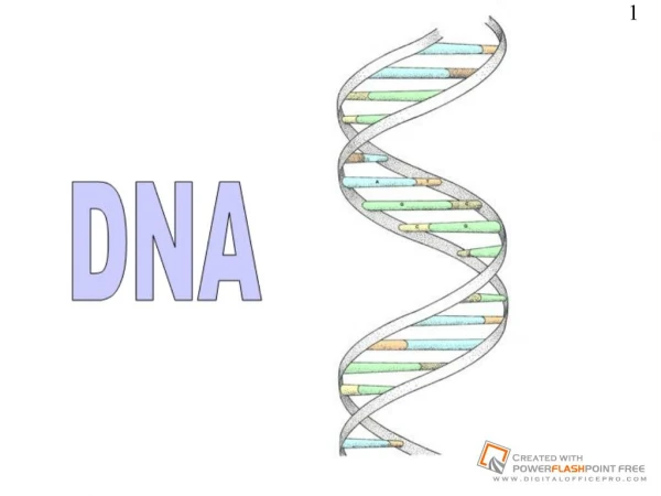
Chromatin Threadlike strands of DNA and histones Arranged in fundamental units called nucleosomes Form condensed, barlike bodies of chromosomes when the nucleus starts to divide Figure 3.29
Cell Cycle Interphase Growth (G1), synthesis (S), growth (G2) Mitotic phase Mitosis and cytokinesis Figure 3.30
Interphase G1 (gap 1) – metabolic activity and vigorous growth G0 – cells that permanently cease dividing S (synthetic) – DNA replication G2 (gap 2) – preparation for division PLAY Late Interphase