Download
1 / 26
260 likes | 567 Vues
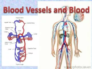
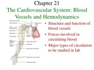
Blood Vessels and Hemodynamics. Chapter 21. Introduction. Blood vessels form a closed system of tubes that carries blood away from the heart, transports it to the tissues and returns it to the heart. Arteries-carry blood away from heart.

E N D
Blood Vessels and Hemodynamics Chapter 21
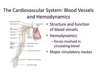
Introduction • Blood vessels form a closed system of tubes that carries blood away from the heart, transports it to the tissues and returns it to the heart. • Arteries-carry blood away from heart. • Divide into small arteries called arterioles which in turn to capillaries. Exchange of substances take place here. • Capillaries unite to form venules which merge to form veins. • Veins then convey blood form the tissues back to the heart. • Larger blood vessels are served by their own blood vessels -vaso vasorum, located within their walls.
Anatomy of Blood Vessels • Arteries carry blood away from the heart. The wall of an artery consists of a tunica interna, a tunica media (which maintains elasticity and contractility) and a tunica externa. • The tunica interna is composed of a lining of simple squamous epithelium (endothelium), a basement membrane and a layer of elastic tissue called internal elastic lamina. Closest to the lumen. • Tunica media-thickest layer. Consists of elastic and smooth muscle fibers. Arteries have high compliance-their walls easily stretch or expand. • Tunica externa-made of elastic and collagen fibers.
Arteries • Sympathetic fibers of the ANS innervate vascular smooth muscle. An inc. in sympathetic stimulation typically stimulates the smooth muscle to contract-vasoconstriction. • When sympathetic stimulation decreases, or in presence of certain chemicals-NO, K+, H+ and lactic acid, they relax-vasodilation. • When an artery or arteriole is damaged, its smooth muscle contracts, producing vascular spasm of the vessel. • This limits blood flow through the damaged vessel and helps reduce blood loss if the vessel is small.
Elastic and Muscular Arteries • Elastic arteries-largest diameter because tunica media contains a high proportion of elastic fibers. They help propel blood onward while the ventricles are relaxing. As blood is ejected their walls stretch. Fn. As pressure reservoir. • They are also termed conducting arteries as they conduct blood from the heart to medium-sized, more muscular arteries.egs. Aorta, brachiocephalic, common carotid, subclavian, vertebral, pulmonary, common iliac.
Muscular Arteries • They are medium-sized arteries. Their tunica media contains more smooth muscle. • Capable of grater vasoconstriction and vaodilation to adjust the blood flow. • Their walls are relatively thick. Also called the distributing arteries. • Egs. Brachial artery in the arm and radial artery in the forearm. • Many of the arteries anastomose ie. The distal ends of two or more vessels unite. An alternate blood route from the anastomosis is called collateral circulation. Those that do not anastomose are the end arteries.
Arterioles • Arterioles are small arteries that deliver blood to capillaries. • Also have the three layers as an artery. • A change in diameter of arterioles can significantly affect blood pressure. • Through constriction and dilation, arterioles assume a key role in regulating blood flow from arteries into capillaries and in altering arterial blood pressure.
Capillaries • These are microscopic blood vessels through which materials are exchanged between blood and tissue cells . • The flowof b lood form arterioles to venules through capillaries is called microcircuilation. • Some capillaries are continuous whereas others are fenestrated. • Capillaries branch to form an extensive network throughout a tissue. • This increases surface area allowing for rapid exchange. • Precappilary sphincters regulate blood flow through capillaries. • Microscopic blood vessels in the liver are called sinusoids.
Capillaries • Capillary walls are made of a single layer of endothelial cells and a basement membrane. They have no tunica media or tunica externa. • Body tissues with high metabolic requirements, such as muscles, kidneys, liver and nervous system, have an extensive network of capillaries. • Tissues with low metabolic requirements have fewer capillaries-tendons and ligaments. • All covering and lining epithelia, cornea and lens of the eye-lack capillaries.
Types of Capillaries • True capillaries:emerge from arterioles and metarterioles. • Continuous capillaries-found in skeletal and smooth muscle, connective tissues and the lungs. • Fenestrated capillaries-kidneys, villi os the SI, choroid plexuses in brain, ciliary process, endocrine glands. • Sinusoids:are wider and more winding than other capillaries. Present in liver, red bone marrow, pleen, ant.pit. Gland, and parathyroid glands.
Veins • Veins consist of the same three tunics as arteries but have a thinner tunica interna and media. The lumen of a vein is also larger than that of an artery. • Veins contain valves to prevent backflow of blood. Weak valves can result in varicose veins. • Vascular sinuses are veins with very thin walls. • Venules are small vessels which are continuous with capillaries and merge to form veins. • Systemic veins are collectively termed blood reservoirs. • The principal blood reservoirs are the veins of the abdominal organs and skin.
Capillary exchange • Substances enter and leave the capillaries by diffusion, vesicular transport (transcytosis) or bulk flow. • The movement of water and solutes (except proteins) through the walls of capillaries depends on hydrostatic and osmotic pressures. • The near equilibrium between filtration and reabsorption in capillaries is called Starling’s law of the capillaries. • Edema is an abnormal increase in interstitial fluid.
Starling’s Law of the Capillaries • Pressure-driven movemnet of fluids and solutes from the blood capillaries into tinterstitial fluid is called filtration, whereas movement from interstitial fluid into capillries is called reabsorption. • Two pressures promote filtration: BHP and IFOP. • The main pressure promoting reabsorption is BCOP. • The balance of the two is called NFP. • Overall, the volume of fluid and solutes normally reabsorbed is almost as large as the volume filtered;this near equilibrium is known as the Sztarling’s Law of the capillaries.
Net Filtration Pressure (NFP) • BHP=35 mmof Hg at arterial end and 16 mm of Hg at venous end. • IFHP=~0 mm Hg • BCOP=26 mm Hg • IFOP=0.1-5 mm Hg. • NFP=(BHP+IFOP)-(BCOP+IFHP) • at arterial end NFP=10 mmHg and hence a net outward pressure and at the venous end NFP=-9 mm Hg which represents a net inward pressure.
Factors affecting Circulation • The velocity of blood flow is inversely proportional to the cross sectional area of the vessels. • The velocity of blood flow decreases form the aorta to arteries to capillaries and increases as blood returns to the heart. • Blood flow is determined by blood pressure and resistance. • Blood flows from regions of higher pressure to lower pressure;higher the resistance, lower the blood flow. • Circulation time is the time required for a drop of blood to pass from the right atrium, through the pulmonary circulation, back to the left atrium, through the systemic circulation down to the foot, and back again to the right atrium. At rest time=1 min.
Blood Pressure • Blood pressure is the pressure exerted on the walls of a blood vessel. • Factors that affect blood pressure are cardiac output, blood volume, viscosity, resistance, and the elasticity of arteries. • As blood leaves the aorta and flows through the systemic circulation, its pressure progressively falls to 0 mm Hg by the time it reaches the right ventricle. • Resistance depends on blood viscosity, blood vessel length, and blood vessel radius.venous return depends on pressure differences between the venules and right ventricle. • Blood return to heart is maintained by several factors.
Blood Pressure and Resistance • MABP=diastolic BP+1/3(sysBP-diasBP) • CO= MABPR • resistance depends on 1)avg.blood vessel radius 2)blood viscosity 3) total blood vessel length. • SVR:systemic vascular resistance
Control of Blood Pressure and Blood Flow • The cardiovascular center (CV) is a group of neurons in the medulla oblongata that regulates heart rate, contractility and blood vessel diameter. • The CV center receives input from higher brain regions and sensory receptors (baroreceptors and chemoreceptors). • Baroreceptors monitor blood pressure and chemoreceptors monitor blood levels of O2, CO2, and hydrogen ions. • The carotid sinus reflex helps regulate blood pressure in the brain. • The aortic reflex is concerned with general systemic blood pressure.
CV center • Output from the CV center flows along sympathetic and parasympathetic fibers. • Sympathetic impulses propagated along cardioaccelarator nerves increase heart rate and contracti9lity, whereas parasympathetic impulses propagated along vagus nerves decrease heart rate. • Hormones that help regulate blood pressure are epinephrine, norepinephrine, ADH (vasopressin), angiotensin II, and ANP. • Autoregulation refers to local, automatic adjustments of blood flow in a given region to meet a particular tissue’s need. • O2 -the principal stimulus for autoregulation.
Shock and Homeostasis • Shock is a failure of the cardiovasculr system to deliver enough O2 to meet the metabolic needs of the cells. • Types of shock include hypovolemic, cardiogenic, vascular and obstructive. • Signs and symptoms include rapid resting heart rate, weak rapid pulse, clammy, cool, pale skin, sweating, hypotension, altered mental state, decreased urinary output, thirst and acidosis.
Evaluating Circulation • Pulse is the alternate expansion and elastic recoil of an artery wall with each heartbeat. It may be felt in any artery that lies near the surface or over a hard tissue. Tachycardia and Bradycardia. • A normal resting pulse (heart) rate is 70-80 bpm. • Blood pressure is the pressure exerted by blood on the wall of an artery when the left ventricle undergoes systole and then diastole. • It is measured using a sphygmomanometer. • Systolic BP is the force of blood recorded during ventricular contraction. Diastolic BP is the force of blood recorded during ventricular relaxation. Normal-120/80. • Pulse pressure-diff bet.sys and dias=40.
Circulatory Routes • The two basic postnatal circulatory routes are the systemic and pulmonary circulations. • Among the subdivisions of the systemic circulation are the coronary (cardiac) circulation and the hepatic portal circulation. • Fetal circulation exists only in the fetus.
Systemic Circulation • The systemic circulation carries oxygenated blood from the left ventricle through the aorta to all parts of the body (including some lung tissue, but not alveoli) and returns the deoxygenated blood to the right atrium. • The aorta is divided into the ascending aorta, the arch of the aorta, and the descending aorta. Each section gives off arteries that branch to supply the whole body. • Blood returns to the heart through the systemic veins. All veins of the systemic circulation drain into the superior or inferior venae cavae or the coronary sinus, which in turn empty into the right atrium.
Hepatic Portal Circulation • The hepatic portal circulation detours venous blood from the GI organs and spleen and directs it into the hepatic portal vein of the liver before it is returned to the heart. • It enables the liver to utilize nutrients and detoxify harmful substances in the blood.
Pulmonary & Fetal Circulation • The pulmonary circulation takes oxygenated blood from the right ventricle to the alveoli within the lungs and returns oxygenated blood from the alveoli to the left atrium. • It allows blood to be oxygenated for systemic circulation. • The fetal circulation involves the exchange of materials between the fetus and mother. • The fetus derives O2 and nutrients and eliminates CO2 and wastes through the maternal blood supply via the placenta.
Aging and the Cardiovascular System • General changes associated with aging include reduced elasticity of blood vessels, reduction in cardiac muscle size, reduced cardiac output, and increased systolic blood pressure. • The incidence of coronary artery disease (CAD), congestive heart failure (CHF) and atherosclerosis increases with age.