Download
1 / 19
190 likes | 417 Vues
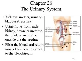
Urinary system كلية الطب البيطري المدرس المساعد حازم كريم ناصر. Urinary system: is subdivided of urogenital system, which has principle function in formation, transport, storage, and excretion of urine. It composed of (k.u.b and u): 1-Right and left kidney (R&L .K)

E N D
Urinary systemكلية الطب البيطري المدرس المساعد حازم كريم ناصر
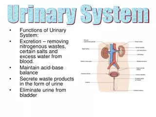
Urinary system: is subdividedof urogenital system, which has principle function in formation, transport, storage, and excretion of urine. • It composed of (k.u.b and u): • 1-Right and left kidney (R&L .K) • 2-Right and left ureter • 3-Urinary bladder. • 4-Urethra
Kidney: they are paired organ present mainly in the sub lumber region, against the dorsal wall of the abdomen. They are retroperitoneal and are partially or completely covered by a fat capsule (depend on the species of animals)
The function of the kidney: • removes waste products from blood (urine); • regulates fluid/salt balance (blood osmotic pressure)
Location of kidney • 1-In pig and cat: the right and left are located in the sub lumber region of the same level. • 2-In horse, dog and camel: right kidney is usually cranial to the left kidney. • 3-In ox: the left kidney is pendulous and present in right side of the abdomen caudal to the right kidney.
Relation of the kidney • 1-Dorsally: left and right kidney (psoas minor, psoas major, and diaphragm) • 2-Ventrally: left and right kidney (intestine and pancreas). • 3-Medially: right kidney (caudal vena), left kidney (abdominal aorta). • 4-Laterally: abdominal wall. • 5-Cranially: left and right kidney related diaphragm. • 6- Caudally: left kidney (spleen), right kidney (liver).
Fixation of the kidney • 1-pressure of adjacent organs. • 2-renal fascia • 3- peritoneum. • 4-ureter. • 5-blood vessels, • 6-renosplenic ligament with left kidney. • 7-renohepatic ligament with right kidney
Classification of kidney • A-According to the cortex (external surface): • 1-Smooth kidney (in all animals except bovine, buffalo, and chicken). • 2-Fissured or furrowed kidney (in bovine , buffalo, and chicken), • B-According to the medulla: • 1- Uni –papillary kidney: in this type the renal papillae are fused to form crest (renal crest) which the common renal papillae is open (in all animals except ox, pig, and man). • 2- Multi papillary kidney: in this type the renal papillae of each lobe is opened separately in small (minor calyx) in ox, pig and man.
C- According to the number of lobules. • 1-Single lobar in mice, rat, and rabbit. • 2-Multi lobular in other mammals has many lobar in each kidney.
Blood supply of kidney: • renal arteries which come from abdominal aorta, branches of these enter at the hilus and on the ventral surface of the kidney and reach the intermediate zone where they form anastomotic arches arcute arteries. From these arcute arteries branches pass into the cortex and medulla. The cortical branches (inter lobular arteries) ) have in general a radial course between the cortical lobules and give off a short lateral branches (perforating and interlobular arteries), each of which ends as the afferent vessels of a renal corpuscle. The blood is carried from the glomerulus by a small efferent vessel, which breaks up immediately into capillaries which form network around the tubules.
The lymph vessels • form net works, capsular or superficial and parenchymaus or deep on leaving the hilus they go to lymph nodes, in this region which are known as the renal lymph nodes.
The nerves supply • The nerves are derived from the renal plexus of the sympathetic, which enclose the renal artery
Ureters • is the narrow portion of the excretory duct of the kidney. Each begins at the renal pelvis and terminates at the urinary bladder. It divided into two parts according to the course: • 1-abdominal portion: each ureter emerges ventrally from the hilus of the kidney and curves caudally and medially toward the lateral face of the caudal vena cava (right side) or the aorta (left side). They then pass almost straight caudally in the psoas minor, cross the external iliac vessels and enter the pelvic cavity. • 2- Pelvic portion: pass caudally and little ventrally on the lateral wall of the pelvic cavity, turns medially, and pierces the dorsal wall of the bladder near the neck.