Download
1 / 18
180 likes | 347 Vues
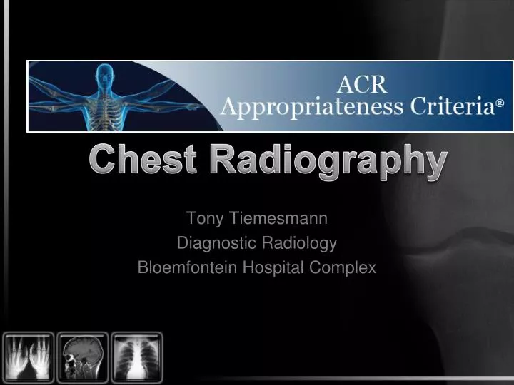
Chest Radiography. Tony Tiemesmann Diagnostic Radiology Bloemfontein Hospital Complex. Introduction. evidence-based guidelines to assist referring physicians in making the most appropriate imaging or treatment decision for a specific clinical condition

E N D
Chest Radiography Tony Tiemesmann Diagnostic Radiology Bloemfontein Hospital Complex
Introduction evidence-based guidelines to assist referring physicians in making the most appropriate imaging or treatment decision for a specific clinical condition enhance quality of care and contribute to the most efficacious use of radiology
ACR Topics • Routine chest radiography • Acute Respiratory Illness in Immunocompetent Patients • Acute Respiratory Illness in Immunocompromised Patients • Chronic Dyspnea — Suspected Pulmonary Origin • Hemoptysis • Non-Invasive Clinical Staging of Bronchogenic Carcinoma • Rib Fractures • Routine Admission and Preoperative Chest Radiography • Routine Chest Radiograph in ICU Patients • Routine Chest Radiographs in Uncomplicated Hypertension • Screening for Pulmonary Metastases • Solitary Pulmonary Nodule
Rating Scale • 1 – 3 • Usually not appropriate • 4 – 6 • May be appropriate • 7 - 10 • Usually appropriate
Routine admission CXR The available evidence does not support a policy for performing routine admission or preoperative chest radiographs for all patients. Although there is no evidence showing that such a policy would lead to worse outcomes for patients, the finding that only 2% of chest radiographs lead to a change in management of patients suggests a high level of cost and inconvenience with potentially limited benefits.
Routine daily CXR in ICU Routine daily chest radiographs are not indicated for patients with unchanged cardiopulmonary problems. In stable patients admitted for cardiac monitoring, or in stable patients admitted for extrathoracic disease only, an initial ICU admission radiograph is not recommended; followup radiographs should be obtained only for specific clinical indications.
Lines/Tubes placement CXR Very few malpositioned tubes are detected by physical examination. Radiographs immediately postintubation are indicated to insure proper positioning. A chest radiographs after insertion of a CVP catheter is recommended to demonstrate proper placement and detect any complications. Beyond the initial insertion, follow-up chest radiographs have a low yield for revealing complications. Follow-up chest radiographs are suggested only when complications are suspected clinically.
Lines/Tubes placement CXR Chest radiographs are suggested after Swan-Ganz catheter insertion. Once pneumothorax has been excluded and proper positioning has been assured, follow-up radiographs are not required except for specific clinical indications. Based on limited evidence, small-bore feeding tubes may, in a small but significant number of patients, be inadvertently placed in the lungs. A chest radiograph is warranted after initial nasogastric tube insertion and before the first feeding. Beyond the initial chest radiograph, follow-up chest radiographs are not required for managing stable tubes.
Lines/Tubes placement CXR After insertion of a chest tube, a chest radiograph is recommended to show the position of the tube, any success in drainage, and possible complications from insertion. Beyond this point, evaluation of tube position and function is warranted based on management of the pleural space and clinical indications.
References American College of Radiology – Appropriateness Criteria reviewed November 2011.