Download
1 / 22
220 likes | 317 Vues
Christina Ziegler Feb 15 th 2010. Mechanisms of tolerance induction. Clonal deletion negative selection of thymocytes with high affinity TCR for MHC:self -antigen (central tolerance) (2) Clonal anergy

E N D
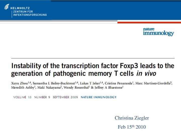
Christina Ziegler Feb 15th 2010
Mechanisms of tolerance induction • Clonal deletion • negative selection of thymocytes with high affinity TCR for MHC:self-antigen (central tolerance) • (2) Clonal anergy • auto-reactive T cells encountering their Ag in absence of co-stimulatory signal become non-responsive to Ag (peripheral tolerance) • (3) Clonal ignorance • removal of auto-reactive T cells not encountering their Ag in periphery • (4) Anti-idiotypic antibody • Ab against specific idiotypes of other Ab or TCR • (5) Regulatory T cells (suppressor cells) • suppressive function via production of TGF-β and IL-10 or cell-cell contact • (6) Termination of tolerance • By prolonged absence/exposure to tolerogen, damage of immune system or immunization with cross-reactive Ag
Development of autoimmune diseases • AUTOIMMUNITY • Breakdown of mechanisms controlling central and/or peripheral tolerance by • Sequestration of antigen • - antigen develops late or is only expressed in particular organ • (2) Escape of autoreactive clones • - defective negative selection in thymus • (3) Lack/deficiency of regulatory T cells • (4) Cross-reactive antigens • - pathogens antigen may cross-react with self-antigens leading to an autoimmune response like e.g. streptococcal nephritis
Characteristics of regulatory T cells Natural Tregs(nTregs) developed in thymus with high affinity for self-antigen - CD25+ Foxp3+ CTLA-4+ (5–10% of total CD4+αβ T cells) Adaptive Tregs(aTregs) develop from conventional T cells in periphery and can be divided into (a) Th3 cells (CD4+ CD25 - Foxp3-) -activated by IL-10 which induced its secretion; acts autocrine (b) Tr1 cells (CD4+ CD25 - Foxp3-) - require IL-10 for maturation, then secrete TGF-β and IL-10 - like Th3 cells, Tr1 are abundant in intestine and likely induce tolerance to food Ag (c) CD8+Tregs(CD8+ CD25 - Foxp3-) - shown to suppress CD4+ cells in vitro
Stability of regulatory T cells – STATUS QUO – • Tregsretain Foxp3 expression under homeostatic conditions after adoptive transfer maybe via positive feedback loop • During inflammation, Tregshave lower Foxp3 expression • Possible that IL-6 acts in synergy with IL-1 to downregulateFoxp3 • CD4+CD25-Foxp3+ were shown to convert into Th cells • SUM: Peripheral Tregscan become unstable under certain conditions.
Mouse model to analyse stability of Tregs Adapted from http://commons.wikimedia.org/wiki/File:CreLoxP_experiment.png
Development of Foxp3+ T cells in Foxp3-GFP-Cre x R26-YFP mice Majority of Foxp3+ cells developed from CD4SP thymocytes (a). Most Foxp3+ transcription is initiated after maturation of CD4SP thymocytes in the thymus (b ). Conclusion: Foxp3+ Treg cells develop as ´escape´ mechanism during negative selection process after exposure to self-Ag.
´Ex-Foxp3´ T cells show fading Foxp3 translation in periphery 15% - 20% of YFP cells lack Foxp3 and GFP expression in thymus and peripheral lymphoid organs, respectively (c). Different peripheral lymphoid organs showed similar proportions of CD4+ T cells expressing Foxp3 at various maturation stages (e). Conclusion: Certain population of T cells called ´ex-Foxp3´ had ceased translation of Foxp3.
Methylation status of ex-Foxp3+ Tregas indicator for their stability Differentiation of Tconv, Tregs and ex-Foxp3 Tregs using CD4 vs Foxp3 or GFP vs YFP (A). Methylation of CpG islands is the principle control mechanism: 90% of CpG motives in TSDR of Foxp3 locus of naive CD4+ Foxp3- Tconv cells are methylated (d). Tregs were mostly de-methylated (GFP+YFP+), while ex-Foxp3 Tregs (GFP-YFP+) Tregs had random methylation status (d). Conclusion: Factors controlling the expression of the Foxp3 led to re-methylation of this locus at certain stage in ex-Foxp3 Tregs .
´Ex-Foxp3´ T cells have a non-Treg cell surface phenotype in the periphery YFP+ ex-Foxp3 T cells were CD25-GITRlowCD127high and thus differ considerably from Foxp3+ Tregs (a). Loss of ´signature´ Treg markers FR4, CTLA-4 and CD103 on ex-Foxp3 T cells in comparison to Tconv and Foxp3 + Tregs (b). Conclusion: Ex-Foxp3 T cells do no longer show Treg specific phenotype indicating their instability in homeostatic conditions. (b) thick line: Tconv cells thin line: Foxp3+ Tregs filled: ex-Foxp3 T cells
´Ex-Foxp3´ T cells show an effector-memory phenotype Ex-Foxp3 T cells (GFP-YFP+) showed an activated-memory T cell phenotype (CD62Llow-highCD44high) (a). Stimulated YFP+ T cells secreted IFN-γ (b) and IL-17 in GALT (c). Th1 or Th17? Conclusion: Ex-Foxp3 T cells show an effector-memory T cell phenotype those cytokine profile depends on the microenviron-ment.
Mouse model to study the Foxp3 expression during an autoimmune disease • NOD MOUSE • the non-obese diabetic mouse is a model of autoimmune disease • develops spontaneous autoimmune diabetes similar to T1D in humans incl. • - pancreas-specific autoantibodies • - autoreactive CD4+ and CD8+ T cells • Inflamed pancreatic β islets have lower Treg to Teffector ratio • Theory: Lower Foxp3 expression in the autoimmune disease shifts balance of Tregs to ex-Foxp3 cell phenotype. • Approach: Crossing of Foxp3-GFP-Cre mouse with R26-YFP-NOD mouse
Autoimmune enviroment favours loss of Foxp3 Fig. legend Panc: pancreas PLN: pancreatic LN ILN: inguinal LN Pancreas contained sig. lower amount of Tregs(GFP+YFP+) but higher percentage of ex-Foxp3 T cells (GFP-YFP+) (a). These ex-Foxp3 T cells were CD25-CD127+ and secreted IFN-γ (b). Conclusion: The autoimmune microenvironment altered the T cell phenotype and promoted pathogenicity. Appearance of ex-Foxp3 T cells was likely consequence of antigen recognition in inflamed area.
Mouse model to study if auto-reactive T cells favour development of ex-Foxp3 T cells • BDC2.5 TCR-tg mouse • TCR of CD4+ T cells in the BDC2.5 TCR-tg mouse are reactive to a natural pancreatic islet β cell antigen • Theory: Auto-reactive T cells in pancreas changes the percentage of ex-Foxp3 cells and their pathogenic potential. • Approach: Crossing of Foxp3-GFP-Cre x R26-YFP mouse with BDC2.5 TCR-tg mouse.
Autoimmune environment favours loss of Foxp3 Proportions of thymic CD4+Tconv and ex-Foxp3 T cells (GFP-YFP+) similar between non-tg and BCD2.5 mice (d). However, spleen and LN of BCD2.5 mice had more ex-Foxp3 cells (d and e) similar to situation in pancreas of NOD mice. Conclusion: Strong affinity to self-antigen especially during inflammation promotes generation of ex-Foxp3 T cells.
Mouse model to study if auto-reactive T cells favour development of ex-Foxp3 T cells NOD Tcra-/- mouse Lack αβT cells and thus are completely protected from autoimmune diabetes. NOD Rag2-/- mouse Has immunodeficiency and combined cellular and humoral immune defects. Theory: Tregs are unstable and potentially pathogenic in autoimmune conditions. Approach: Adoptive transfer of Tregs from Foxp3-GFP-Cre x R26-YFP x BDC2.5 TCR-tg mouse into a) NOD Tcra-/- mouse and b) NOD Rag2-/- mouse
Ex-Foxp3 cells can be generated from nTregs or aTregs Adoptively transferred nTregsfrom BDC2.5 TCR-tg Foxp3-GFP-Cre x R26-YFP mouse into the NOD Tcra-/- a) had to 1/3 down-regulated Foxp3, b) effector-memoryphenotype(a). After adoptive transfer of Foxp3- cells into the NOD Rag2-/- mouse, thoseexpressing BDC2.5 TCR were 0.3% YFP+ in thepancreas. Conclusion: Ex-Foxp3 cells can be generated from instable nTregs or to a lesser extend from abortive aTregs.
Auto-reactive ex-Foxp3 T cells turn into effector cells and then induce T1D • Ex vivo expansion of ex-Foxp3, Tconv and Tregs from BDC2.5 TCR-tg mice for 7-9 d. • 20% of GFP+YFP+ and 2% of YFP+ lost Foxp3 expression (b). • Adoptive transfer of three T cell subtypes into the NOD Rag2-/- mouse • Tregs did not alter the blood glucose levels • Tconvand ex-Foxp3 T cells induced diabetes (c and d). • Conclusion: Auto-reactive Ex-Foxp3 T cells turn into effector cells after self-antigen recognition and induce T1D.
Ontogeny of ex-Foxp3 T cells Unclear if ex-Foxp3 originate from i) aborted Fopx3+aTreg cells that had converted from Tconv or ii) Tconv in the periphery or iii) loss of Foxp3 expression in true CD4+Foxp3+nTreg cells Analysis of the CDR3 in various CD4+ T cell subsets from BDC2.5 TCR-tg mice showed that i) all subsets had productive VJ gene rearrangement ii) Treg and Tconvcells had dinstinct TCR Vα2 repertoire as only 13% of CDR3 sequence was present in Tconv iii) Ex-Foxp3 cells shared 24% and 36 % sequence CDR3 similarity to Treg and Tconv, respectively. Conclusion: Ex-Foxp3 cells have substantial overlap of TCR repertoire with Treg and Tconv and can probably originate from both T cell subtypes.
Summary and conclusions • Substantial fraction of Tregs are unstable in the periphery as a significant percentage • down-regulates Foxp3 • loses their characteristic Treg phenotype • exhibits an activated-memory phenotype and • produces pathogenic cytokines • loses their suppressive function • triggers development of autoimmune disease • ´ex-Foxp3´ T cell levels were elevated in autoimmune conditions • cells share ontogeny with Foxp3+Tregs and Tconvthus likely originate from nTregs and aTregs • THEORY: Foxp3 instability can lead to the generation of pathogenic effector-memory T cells that promote autoimmunity.
Possible reasons for the development of autoimmune diseases • Foxp3 instability can lead to the generation of pathogenic effector-memory T cells that promoter autoimmunity • Functional deficiency of IL-2 signalling in Treg cells in autoimmunity may disturb the positive feedback loop that controls Foxp3 stability • Dysfunctional microRNA or Dicer can affect Foxp3 stability • Destabilized Foxp3 possibly involves epigenetic changes in the Foxp3 locus • Early inflammatory cytokines induced by the innate immune system may disable Tregs and enhance immunity by creating locally pathogenic autoreactive T cell repertoire