Download
1 / 23
260 likes | 659 Vues
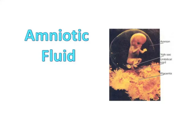
Amniotic F luid Embolism. Dr. Megha jain. University College of Medical Sciences & GTB Hospital, Delhi. www.anaesthesia.co.in. email: anaesthesia.co.in@gmail.com. Amniotic fluid embolism. Occurs when amniotic fluid, fetal cells, hair or other debris enter maternal circulation.

E N D
Amniotic Fluid Embolism Dr. Meghajain University College of Medical Sciences & GTB Hospital, Delhi www.anaesthesia.co.in email: anaesthesia.co.in@gmail.com
Amniotic fluid embolism Occurs when amniotic fluid, fetal cells, hair or other debris enter maternal circulation. during labour during caesarean section after normal vaginal delivery during II trimester termination of pregnancy 1 in 8000 to 80,000 pregnancies. Definition Timing Incidence
Risk factors • Advanced maternal age • Multiparity • Meconium stained liquor • Placenta accreta • Polyhydramnios • Uterine rupture • Cervical laceration • Strong uterine contractions • Chorioamnionitis • Maternal atopy/ allergy
Pathophysiology Phase 1 Debris in maternal circulation Biochemical mediators (endothelin,LT’s) Pulmonary artery spasm (pulm HTN) RV pressure Hypoxia Myocardial damage LVF ARDS
Pathophysiology (contd….) Biochemical mediators Activation of factor X DIC (30min to 4 hr after phase 1) Hemorrhagic phase Massive hemorrhage & uterine atony Phase 2
Immunological response to amniotic fluid exposure Maternal exposure to fetal tissue Majority patients – No effect Small no. of patients- Endogenous mediator release S/S depends on antigenic exposure And individual response SEVERE Hypoxia Cardiovascular collapse Coagulopathy Death LESS SEVERE: isolated finding Or prolonged presentation
Classical Triad ACUTE HYPOXIA HEMODYNAMIC COAGULOPATHY COLLAPSE
Signs and Symptoms • Hypotension • Fetal distress • Pulmonary edema or ARDS • Cardiopulmonary arrest • Cyanosis • Coagulopathy • Dyspnea • Seizure • Uterine atony • Bronchospasm • Chest pain, dysrhythmias
Is AFE an anaphylactoid reaction? • Previous history of drug allergy or atopy • Mediators assoc. with anaphylactoid reaction are released in AFE. • AFE is not due to physical obstruction of pulmonary vasculature • Pre treatment with LT inhibitor prevents AFE collapse • Treatment with antihistaminic reduced degree of shock • Steroid - successfully used in mgmt. of AFE.
How to diagnose ? • NON SPECIFIC • Complete blood count • FDP, Fibinogen • Coagulation profile • ABG • CXR • ECG • V/Q scan • SPECIFIC • Zinc Coproporphyrin • > 35 nmol/L • in maternal plasma • (a component • of meconium) • 2. Serum tryptase -
Lab tests in DIC • Fibrinogen < 150mg/dl 400-650 • Plt. Count < 50,000/mm3 1.5-3 lacs • Thrombin time> 100 sec 15-20 sec • PT > 100 sec 10-12 sec • PTTK > 100 sec 35-50 sec • FDP > 200 µg/ml < 16 µg/ml DIC Normal values
Differential diagnosis 1. Cardiovascular – Myocardial infarction Primary arrhythmia Hemorrhage(APH,PPH) 2. Respiratory - Pulmonary embolism Air embolism Aspiration pneumonitis 3. CNS - Eclampsia 4. Regional - High/ Total spinal anesthesia LA toxicity 5. Anaphylactoid reaction 6. Septic shock
Management GOALS • Restore CVS and pulmonary equilibrium • Maintain SBP > 90 mmHg • UO > 25 ml / hr • Arterial PO2 > 60 mmHg • Re establishing uterine tone • Correct coagulation parameters
Immediate measures Convert regional to GA immediately # Intubate and ventilate with 100% oxygen # Initiate CPR if indicated # If undelivered, monitor fetus and deliver # Provide aggressive volume and pressor support pressor support may include- *Dopamine: 2 – 5 µg/kg/min *Dobutamine: 15 – 30 µg/kg/min *Noradrenaline: 0.1 – 0.4 µg/kg/min *Adrenaline: 0.15 – 0.30 µg/kg/min # Restrict fluid to maintainence levels ( ARDS follows in 70%)
Restore uterine tone Uterine massage Uterine packing Oxytocin PG analouges – PG F2 alpha Improve CO & Uterine perfusion Displacement of the patient’s uterus towards left – improves venous return and uterine blood flow.
Manage coagulopathy DIC – depletion of fibrinogen, platelets, coagulation factors Invg- PT/ PTTK, platelet count Treatment 1. FFP 2. cryoprecipitate – fibrinogen <100 mg/dl 3. platelet transfusion – at < 50,000 / mm3 or if spontaneous bleeding is present.
Management in ICU Monitor – ECG, PO2, PCO2, urine output Arterial catheterization – for repeated ABGs CV catheterisation – diagnose RV overload guide fluid therapy Pulmonary artery catheterisation and PCWP- measure LV function and compliance
Prognosis Maternal 60% die within 1 hr of embolism Of survivors, 75% have long term neurological deficits 50% develop DIC (persistent bleeding) 10-15% develop GTCS Mortality – 1. sudden cardiac arrest 2. hemorrhage ( coagulopathy) 3. ARDS and multiple organ failure (if in utero at the time of event) 70% survive, 50% have neurological deficit. Fetal
Thank You www.anaesthesia.co.in