Download
1 / 46
810 likes | 1.75k Vues
Muscle tissue. Larry Johnson Texas A&M University. Objectives. Histologically identify and functionally characterize each of the 3 types of muscle tissues. Describe the organization of the sarcomere as seen in light and electron microscopy.

E N D
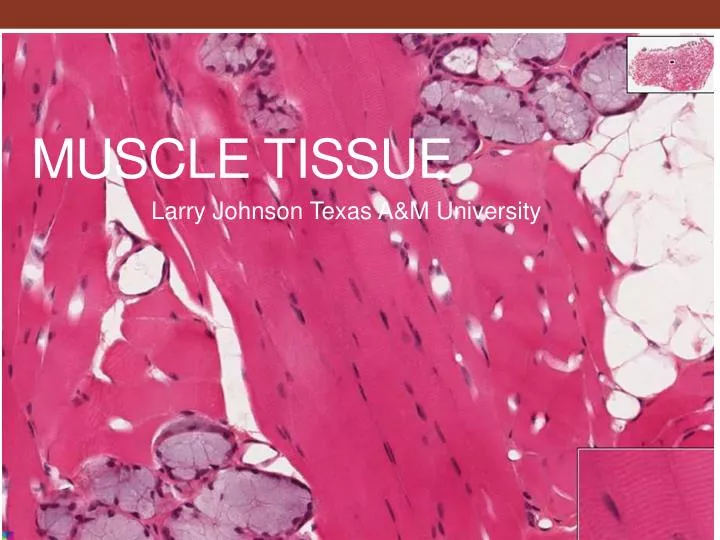
Muscle tissue Larry Johnson Texas A&M University
Objectives • Histologically identify and functionally characterize each of the 3 types of muscle tissues. • Describe the organization of the sarcomere as seen in light and electron microscopy. • Identify the endomysium, perimysium, and epimysium CT sleeves in muscle. • Relate the functional differences of the three muscle cell types. From: Douglas P. Dohrman and TAMHSC Faculty 2012 Structure and Function of Human Organ Systems, Histology Laboratory Manual
MUSCLE FUNCTION: • GENERATION OF CONTRACTILE FORCE DISTINGUISHING FEATURES: • HIGH CONCENTRATION OF CONTRACTILE PROTEINS ACTIN AND MYOSIN ARRANGED EITHER DIFFUSELY IN THE CYTOPLASM (SMOOTH MUSCLE) OR IN REGULAR REPEATING UNITS CALLED SARCOMERES(STRIATED MUSCLES, e.g., CARDIAC AND SKELETAL MUSCLES)
MUSCLE • DISTRIBUTION: SKELETAL – STRIATED MUSCLES MOSTLY ASSOCIATED WITH THE SKELETON CARDIAC – STRIATED MUSCLES ASSOCIATEWD WITH THE HEART SMOOTH – FUSIFORM CELLS ASSOCIATED WITH THE VISCERA, RESPIRATORY TRACT, BLOOD VESSELS, UTERUS, ETC.
MUSCLE • HISTOLOGICAL INDENTIFICATION: SKELETAL MUSCLE – VERY LONG CYLINDRICAL STRIATED MUSCLE CELLS WITH MULTIPLE PERIPHERAL NUCLEI CARDIAC MUSCLE – SHORT BRANCHING STRIATED MUSCLE CELLS WITH CENTRALLY LOCATED NUCLEI SMOOTH MUSCLE – CLOSELY PACKED SPINDLE-SHAPED CELLS WITH A SINGLE CENTRALLY PLACED NUCLEUS AND CYTOPLASM THAT APPEARS HOMOGENEOUS BY LIGHT MICROSCOPY Myoepithelial cells
Muscle Tissue • Muscle cells specialized for contraction with apparatus of actin and myosin proteins. • 3 types:
Slide 11: Skeletal muscle (longitudinal and cross sections) Muscle fibers with peripheral nuclei Cross sections of muscle fibers with peripheral nuclei Capillary
Slide 052: Skeletal muscle (longitudinal and cross sections) Cross striations (A and I bands) Muscle fibers with peripheral nuclei Capillary The increase in muscle mass during exercise results from stimulating formation of new myofibrils and enlargement in the diameter of individual muscle fibers (hypertrophy = increase in size).
CONNECTIVE TISSUE LAYERS OF SKELETAL MUSCLE • EPIMYSIUM - COARSE CT • PERIMYSIUM - LESS COARSE CT • ENDOMYSIUM - DELICATE CT PERIMYSIUM EPIMYSIUM ENDOMYSIUM
CONNECTIVE TISSUE LAYERS OF SKELETAL MUSCLE EPIMYSIUM, PERIMYSIUM, and ENDOMYSIUM
Slide 11: Skeletal muscle (longitudinal and cross sections) Epimysium Perimysium Endomysium
Slide 052 Skeletal muscle (longitudinal and cross sections) Perimysium Endomysium with its capillaries
CONNECTIVE TISSUEconnect LAYERS OF SKELETAL MUSCLE • ENDOMYSIUM
CONNECTIVE TISSUE LAYERS OF SKELETAL MUSCLE PERIMYSIUM • ENDOMYSIUM 052
Striations in skeletal muscle 052 Skeletal muscles have striations, peripheral nuclei, same thickness throughout their length and do not branch. Specialized for powerful and rapid contraction. Voluntary. Found attached to the skeleton.
Slide 12: Skeletal muscle (phosphotungstic acid/hematoxylin) Sarcomere: Z line to Z line Z line is In the I band I band A band
STRIATED MUSCLE • CARDIAC A I SKELETAL “A” BAND = dark band ANISOTROPIC = DOES ALTER POLARIZED LIGHT (BIREFRINGENT) “I” BAND = light band ISOTROPIC = DOES NOT ALTER POLARIZED LIGHT
STRIATED MUSCLE (SKELETAL) A I SARCOMERES ARE ORGANIZED FOR RAPID AND HIGHLY CONTROLLED CONTRACTION
INTERMEDIATE FILAMENTS – FUNCTION in muscle cells • MYOFIBRIL ORGANIZATION – MUSCLE cells Cell =
CONTRACTION OF THE SARCOMERE • THIN FILAMENT ACTIN (F-ACTIN) TROPOMYOSIN TROPONIN T - ATTACHES TO TROPOMYOSIN C - BINDS CALCIUM IONS I - INHIBITS ACTIN-MYOSIN INTERACTION • The stoppage of the neural impulse and depletion of free calcium ends the actin-myosin crossbridge cycle in skeletal muscle. ATP is required in this process. • .
CALCIUM REGULATION • TRANVERSE (T) TUBULE (INVAGINATION OF SARCOLEMMA) • TRANSMIT DEPOLARIZATION OF MEMBRANE DEEP INTO THE CELL • SARCOPLASMIC RETICULUM (SER OF CELL) RELEASE CA++ FOR CONTRACTION - THEN RECOVER CA++ • AFTER CONTRACTION • TRIAD = (T TUBULE AND TWO ENDS OF SER)
Slide 13: Skeletal Muscle – muscle spindles (trichrome) Intrafusal muscle fibers Muscle spindle: (stretch receptors) Nerve fibers
INNERVATION OF MUSCLE MUSCLE SPINDLE MUSCLE SPINDLE Intracapsular fibers
SMOOTH MUSCLE Has a PAS + basement membrane
Slide 35: Urinary bladder Muscularis externa (smooth muscle) Smooth muscle fibers with central nuclei Smooth muscles are fusiform with tapered ends, single, central nuclei, and no striations. Involuntary. Found in sphincters and sheets of internal visceral organs and glands.
Slide 63: Appendix Muscularis externa Submucosa Smooth muscle layer arrangement in tubular organs The layering of smooth muscle in the gut is responsible for contractions which mix and propel luminal contents forward.
EM 49: Smooth muscle Actin Myosin
Skeletal Muscle Smooth muscle
CARDIAC MUSCLE IS STRIATED MUSCLE • INTERCALATED DISC • FASCIA ADHERENS • MACULAE ADHERENS • GAP JUNCTIONS - LATERAL PORTION
CARDIAC MUSCLE INTERCALATED DISC The intercalated discs enables coordinated function via gap junctions to facilitate energy and calcium conductance between neighboring myocytes.
pulmonary artery in the lungs of a rat INTERCALATED DISC nucleus mitochondria
Slide 14: Heart & EM 48: Heart Intercalated discs
CARDIAC MUSCLE – Diad located at Z line Diad = (T tubule + one end of SER)
REGENERATION OF MUSCLE Which type of muscle cells can be replaced in adults? • SMOOTH – LOTS • SKELETAL – SOME • CARDIAC – NONE
Clinical Correlation Ischemic heart disease is one of the most significant health problems in the US. Coronary artery thrombosis usually precedes and precipitates a myocardial infarct, resulting in the death of cardiac myocytes. Eventually scar tissue is formed and there is some loss of contractility. Image taken from adultstemcellawareness.wordpress.com Lacking muscle mesenchymal satellite cells, adult mammalian cardiac muscle has little potential to regenerate after injury. Damage is replaced by proliferating fibroblasts and connective tissue growth, forming myocardial scars.
Many illustrations in these VIBS Histology YouTube videos were modified from the following books and sources: Many thanks to original sources! • Bruce Alberts, et al. 1983. Molecular Biology of the Cell. Garland Publishing, Inc., New York, NY. • Bruce Alberts, et al. 1994. Molecular Biology of the Cell. Garland Publishing, Inc., New York, NY. • William J. Banks, 1981. Applied Veterinary Histology. Williams and Wilkins, Los Angeles, CA. • Hans Elias, et al. 1978. Histology and Human Microanatomy. John Wiley and Sons, New York, NY. • Don W. Fawcett. 1986. Bloom and Fawcett. A textbook of histology. W. B. Saunders Company, Philadelphia, PA. • Don W. Fawcett. 1994. Bloom and Fawcett. A textbook of histology. Chapman and Hall, New York, NY. • Arthur W. Ham and David H. Cormack. 1979. Histology. J. S. Lippincott Company, Philadelphia, PA. • Luis C. Junqueira, et al. 1983. Basic Histology. Lange Medical Publications, Los Altos, CA. • L. Carlos Junqueira, et al. 1995. Basic Histology. Appleton and Lange, Norwalk, CT. • L.L. Langley, et al. 1974. Dynamic Anatomy and Physiology. McGraw-Hill Book Company, New York, NY. • W.W. Tuttle and Byron A. Schottelius. 1969. Textbook of Physiology. The C. V. Mosby Company, St. Louis, MO. • Leon Weiss. 1977. Histology Cell and Tissue Biology. Elsevier Biomedical, New York, NY. • Leon Weiss and Roy O. Greep. 1977. Histology. McGraw-Hill Book Company, New York, NY. • Nature (http://www.nature.com), Vol. 414:88,2001. • A.L. Mescher 2013 Junqueira’s Basis Histology text and atlas, 13th ed. McGraw • Douglas P. Dohrman and TAMHSC Faculty 2012 Structure and Function of Human Organ Systems, Histology Laboratory Manual - Slide selections were largely based on this manual for first year medical students at TAMHSC