Download
1 / 78
790 likes | 800 Vues
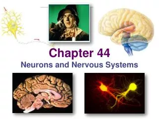
Nervous System - Neurons. Biol 105 Lecture 9 Chapter 7. Outline. Nervous system function Central and peripheral nervous system Nervous system cells Myelinated neurons Nerve signal transmission Nerve Synapse. Nervous Tissues.

E N D
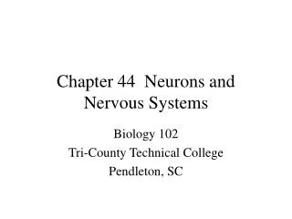
Nervous System - Neurons Biol 105 Lecture 9 Chapter 7
Outline • Nervous system function • Central and peripheral nervous system • Nervous system cells • Myelinated neurons • Nerve signal transmission • Nerve Synapse
Nervous Tissues • Nervous tissue functions to conduct messages throughout the body. • When nerve cells are stimulated, an electrical signal quickly travels through the nerve cell to the nerve ending, triggering events
Nervous System • Includes nerve tissue and sense organs • Nervous system functions to: • Senses environment – receives information from both outside and inside the body • Processes the information it receives • Respond to information – sends out orders
Two Parts of the Nervous System • Central Nervous System (CNS) • Brain and Spinal Cord • Peripheral Nervous System (PNS) • Nervous tissue outside brain and spine • Sense organs
Central Nervous System Peripheral
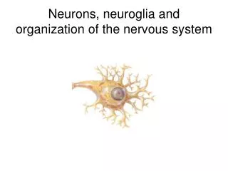
Nervous System Cells • Two types of nervous tissue cells • Neurons – the cells that are responsible for transmitting messages • Neuroglial cells – cells that support the neurons
Neuroglial cells • Microglia – immune system cells, engulf bacteria and cellular debris • Astrocytes – provide nutrients to neurons • Oligodenrocytes and Schwann cells – form myelin sheaths
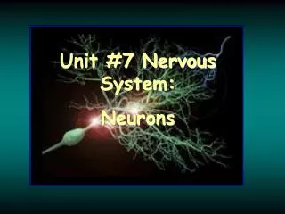
Parts of a Neuron • Cell body – contains the nucleus, main body of cell • Dendrites – projections from the cell body that carry messagesto the cell body • Axons – one large projection that carry messagesaway from the cell body
Neurons Have Dendrites, a Cell Body, and an Axon Dendrites receive information from other neurons or from the environment. The cell body controls the cell’s metabolic activities. Axon endings release chemicals called neurotransmitters that affect the activity of nearby neurons or an effector (muscle or gland). Nucleus Cell body An axon conducts the nerve impulse away from the cell body. The cell body integrates input from other neurons. Axon endings Sending portion of neuron Receiving portion of neuron Figure 7.2
Neurons of the Peripheral Nervous System • Neurons in the PNS are either carrying messages to or from the CNS • Afferent = Sensory neurons = Neurons carrying messages to the CNS • Efferent = Motor neurons = Neurons carrying messages from the CNS
Interneurons in the Central Nervous System • Located between sensory and motor neurons within the CNS • Interneurons integrate and interpret sensory signals
Sensory Neurons • The afferent or sensory neuron cell bodies are located in dorsal root ganglion.
Motor Neurons • The efferent or motor neuron cell bodies are located in the gray matter of the spinal cord. • Their axons leave the CNS and go to the skeletal muscles
The cell bodies of these neurons are located in the dorsal root ganglion • Motor • Sensory
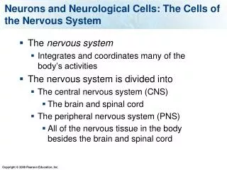
Neurons of the Nervous System Muscle (effector) Sensory receptor for pain Impulse direction Sensory neuron Cell body Motor neuron Interneuron Figure 7.1
These neuroglial cells provide nutrients to neurons • Microglia • Astrocytes • Oligodenrocytes • Schwann cells
These are projections of the neuron cell body that carry messagesto the cell body • Axons • Dendrites
Which of the following type of neuron would alert the brain that you had touched a hot object? • efferent neuron • afferent neuron
What type of neuron is the arrow pointing to? • Sensory • Motor
Myelinated neurons • Neurons that have axons covered with neuroglial cells that contain the protein myelin are called myelinated neurons • Myelinated neurons are able to carry messages faster than non-myelinated neurons
Functions of Myelin Sheaths • The main benefit of myelin sheaths is that myelinated neurons are able to carry messages faster than non-myelinated neurons • Myelin sheaths from Schwann cells also help regenerate injured PNS neuron axons
Two Types of Cells that Myelinate neurons • Schwann cells and Oligodenrocytes are wrapped around neuronal axons
Myelinated neurons • Schwann cells are found in the PNS • Oligodendrocytes are found in the CNS • Nodes of Ranvier are spaces on the axon between the glial cells
Myelinated Neurons http://www.youtube.com/watch?v=mOgHC5G8LuI
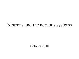
Myelinated Neurons Nucleus Dendrites Cell body In saltatory conduction, the nerve impulses jump from one node of Ranvier to the next. Node of Ranvier Schwann cell Myelin sheath (a) Figure 7.3a
Myelin Sheath Figure 7.3b
Myelin Sheath Figure 7.3c
Multiple Sclerosis (MS) • Caused by the destruction of the myelin sheath that surrounds axons found in the CNS • Can result in paralysis and loss of sensation, including loss of vision
Nerve • Nerve contain Neuron axons are bundled together • These bundles contain • Axons • Blood vessels • Connective tissue
Nerve Connective tissue surrounding one nerve Blood supply Axons within a connective tissue sheath One axon (d) The anatomy of a nerve Figure 8.9d
An Ion is an atom that has gained or lost a • Neutron • Proton • Electron
How can an ion pass through a membrane • Simple diffusion • Facilitated diffusion • Active transport • Both 2 and 3 • All of the above
The Nerve Impulse Is an Electrochemical Signal • A nerve impulse, or action potential, involves sodium ions (Na+) and potassium ions (K+) that cross the cell membrane through the ion channels • Each ion channel is designed to allow only certain ions to pass through
Action Potential Cross section Axon membrane Neuron plasma membrane Extracellular fluid Cytoplasm Continually open ion channels Sodium-potassium pump “Gated” ion channels Sodium-potassium pump The sodium-potassium pump uses cellular energy (ATP) to pump sodium ions out of the cell and potassium ions into the cell Ion channels Ion channels can be open continuously or opened and closed by a molecular gate Figure 7.4
Membrane Potential • The difference in charge between the inside and outside of the neuron is the membrane potential
Resting Membrane Potential • A neuron that is not conducting a message is said to be “Resting” • When a neuron is resting there is more sodium (Na+) outside the neuron cell and more potassium (K+) inside the cell • The inside of the cell has a negative charge compared to the outside the cell
The Nerve Impulse Figure 7.5 (1 of 4)
Sodium Potassium Pump • To maintain this resting membrane potential the neuron pumps Na+ out of the cell and K+ into the cell. • The transport proteins take 3 Na+ ions out for every 2 K+ ions into the cell = Na+/K+ pump • This is Active Transport – requiring ATP
Action Potential • An electrochemical signal conducted along an axon. It is a wave of depolarization followed by repolarization • Depolarization is caused by sodium ions entering the axon • Repolarization is caused by potassium ions leaving axon
Steps of an Action Potential • The axon is depolarized when voltage gated sodium ion channels open and Na+ comes rushing in, causing the inside of the neuron to become positively charged
Action Potential Figure 7.5 (2 of 4)