Download
1 / 38
400 likes | 648 Vues
DNA. First isolated from the nuclei of cells in 1869 Oswald Avery (1944) presented evidence that suggested that nucleic acids were involved in the storage and transfer of genetic information. Erwin Chargaff found that the DNA always contains the same relative amounts of certain pairs of amine ba

E N D
1. DNA and the Genetic Code Larry J. Scheffler
Lincoln High School
2009
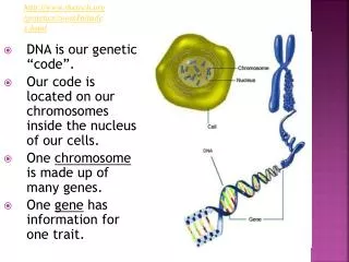
2. DNA First isolated from the nuclei of cells in 1869
Oswald Avery (1944) presented evidence that suggested that nucleic acids were involved in the storage and transfer of genetic information.
Erwin Chargaff found that the DNA always contains the same relative amounts of certain pairs of amine bases. There are always equal amounts of
adenine and thymine.
guanine and cytosine.
James Watson and Francis Crick in
1953 determined the structure of DNA
as a double helix.
Rosalind Franklin created the early X-ray diffraction pictures of DNA.
3. Nucleic Acids Nucleic acids fall into two classes,
-- DNA
-- RNA
RNA, or Ribonucleic Acid, is built on the �-D-ribofuranose ring.
DNA, or deoxyribonucleic acid, is based on a modified ribofuranose ring in which the -OH group on the second carbon atom has been removed.
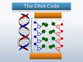
4. DNA Structure DNA is made up of three units including:
I. A ribose sugar
5. DNA Structure DNA is made up of three units including
I. A ribose sugar
II. A phosphate group
6. DNA Structure DNA is made up of three units including
I. A ribose sugar
II. A phosphate group
III. A nitrogen (amine) base
7. A Nucleotide The DNA strand is made up of alternating deoxyribose and phosphate groups with a nitrogen base attached as a side chain.
8. DNA Structure DNA is made up of three units
A ribose sugar
A phosphate group
A nitrogen (amine) base
9. DNA Structure DNA is made up of three units:
A ribose sugar
A phosphate group
A nitrogen (amine) base
These three molecules make up a nucleotide.
A DNA strand is a sequence of nucleotides.
The phosphates are attached at carbon 3 and carbon 5.
The nitrogen bases are side chains at carbon 1.
10. DNA Structure DNA consists of two strands of nucleotides. These strands are wound together in a spiral known as a double helix
The amine bases hold the strand together with a
sequence
of hydrogen
bonds
11. Complimentary Bases Because of their size and their ability to hydrogen bond, the amine bases exist in complimentary pairs in the DNA double helix
Adenine always bonds with Thymine and Guanine always bonds with Cytosine
12. Hydrogen Bond Alignment The size and shape of the amine bases is such that hydrogen bonds can only form at specific sites
Adenine only bonds with Thymine
Guanine only bonds with Cytosine
Therefore they form complimentary base pairs
13. DNA Structure -- Hydrogen Bonding Adenine and Thymine form a base pair
14. DNA Structure Hydrogen Bonding Guanine and Cytosine
15. Base Pair Sequence The sequences of bases appears to be random but in reality nothing is farther from the truth. The base pair sequence contains the code by which proteins are synthesized in the cell
16. DNA Structure In the double helix of a DNA molecule, the two strands are not parallel, but interwoven with each other.�
The helix makes a turn every 3.4 nm, and the distance between two neighboring base pairs is 0.34 nm.�
There are about 10 pairs per turn.�
The intertwined strands make two grooves of different widths, known as the major groove and the minor groove.
These grooves may facilitate binding with specific proteins.
17. DNA Shape This color enhanced image taken by the Scanning Tunneling Electron Microscope shows a double helix
18. DNA Replication In human beings there are 23 pairs of chromosomes
Chromosomes are effectively a very long DNA sequence. This DNA sequence replicates itself during cell division
As the DNA double helix partially unzips as the hydrogen bonds between the nitrogen bases are broken
Sugar and base units are picked up from the surrounding solution.
Since only A �T and G-C combinations can occur the new strand is a complimentary replicate of the existing DNA
19. DNA Replication When cells divide the DNA must is replicated exactly
As the DNA unzips new complimentary strands are formed.
These new strands are exact replicas of the previously existing strands
20. DNA and the Genetic Code Genes are long sequences of DNA that code for the formation of proteins
Typical genes are often thousands of base pairs long
Not all of the DNA strand appears to have genetic information
The sequence for a particular gene is very specific.
21. Gene Correspondance for Neuropilin-1
22. Protein Synthesis DNA is found in the chromosomes which are found in the nucleus of the cell
DNA stores the genetic code for an organism through its sequence of the nitrogen bases
The genetic code is transferred via RNA to the ribosomes in the cytoplasm outside of the cell nucleus where protein in synthesized
The information required for protein synthesis is passed through a similar unzipping and replication process
23. RNA and Protein Synthesis The transfer of information for building proteins is then accomplished by the RNA.
RNA is similar to DNA but there are some important differences
RNA is a single strand rather than a double helix
Deoxyribose is replaced with ribose
The nitrogen base thymine is replaced with tracil
24. RNA Structure Ribose has a slightly different structure from deoxyribose.
Ribose has an �OH group on carbon 2 rather than a H as in deoxyribose.
25. RNA v DNA The structure of Uracil differs only slightly from Thymine
26. Messenger RNA Messenger RNA or mRNA copies and carries the genetic code from the DNA template within the cell nucleus to the ribosomes where proteins are synthesized.
It essentially aligns itself with the DNA and produces a complimentary copy
27. Messenger RNA Messenger RNA (mRNA) acts as a template for protein synthesis
It has the same sequence of bases (in the 5' to the 3' direction) as the DNA strand that holds the gene sequence.
mRNA strands can range from 300 to as many as 7000 nucleotides.
The length depends on the size and the number of proteins related to the code.
28. Transfer RNA Transfer RNA acts as an amino acid carrier in the formation of proteins.
Through a decoding mechanism it facilitates the addition of an amino acid to a peptide chain forming a protein.
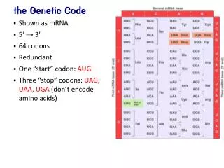
It directs the insertion of amino acids in the proper sequence in the poly peptide chain through sets of three nitrogen bases known as codons.
29. Transfer RNA tRNA molecules are covalently attached to the corresponding amino-acid at one end, and at the other end they have a triplet sequence (called the anti-codon) that is complementary to the triplet codon on the mRNA.
All tRNA molecules are in the range ~70-90 nucleotides. They have a molecular weight of ~25,000
30. RNA Codons
31. Ribosomal RNA Ribosomal RNA (rRNA) is one of the structural components of a cell structure known as a Ribosome.
Ribosomes structurally support and catalyze protein synthesis.
32. DNA Replication During Cell Division During cell replication the DNA unwinds and each strand builds a new complimentary strand.
33. RT-PCR or Reverse Transcriptase Polymerase Chain Reactions
34. RT-PCR and DNA Replication Semi-quantitative RT-PCR was used to ascertain the relative presence of neuropilin-1 in assays from various stages of the menstrual cycle. Cyclophilin was used as an internal standard.
The NP-1 band confirms the presence of neuropilin-1 at all luteal phases. The relative intensity of the NP-1 band relative to cyclophilin appears to be elevated at thre midlate stage.Semi-quantitative RT-PCR was used to ascertain the relative presence of neuropilin-1 in assays from various stages of the menstrual cycle. Cyclophilin was used as an internal standard.
The NP-1 band confirms the presence of neuropilin-1 at all luteal phases. The relative intensity of the NP-1 band relative to cyclophilin appears to be elevated at thre midlate stage.
35. DNA Sequencing
36. Forensic DNA Analysis
37. DNA Fingerprinting Process DNA fingerprinting is a multistep process.
38. Forensic DNA Analysis
39. Forensic DNA Analysis