Download
1 / 60
670 likes | 1.49k Vues
FRACTURES. PEDIATRIC EMERGENCY DEPARTMENT COHEN CHILDREN’S MEDICAL CENTER. FRACTURES. OUTLINE. Statistics Definitions Anatomy Clinical Presentation Diagnosis Management Clinical Scenarios. Statistics. Skeletal injuries account for 10-15% of injuries in children

E N D
FRACTURES PEDIATRIC EMERGENCY DEPARTMENT COHEN CHILDREN’S MEDICAL CENTER FRACTURES
OUTLINE • Statistics • Definitions • Anatomy • Clinical Presentation • Diagnosis • Management • Clinical Scenarios
Statistics • Skeletal injuries account for 10-15% of injuries in children • 15% of these injuries affect the growth plate • The number and spectrum of musculoskeletal injuries in pediatrics is on the rise due to more participation by youth in high risk sports and activities. • The clavicle, radius, and ulna are the most frequently fractured bones in children
Definitions • Fracture: A break in the continuity or architecture of a bone • Open Fracture: communicates with the outside environment via puncture or laceration • Pathologic Fracture: occurs in areas of bone weakness (rickets, bone cysts, osteogenesis imperfecta, malignancies)
Definitions • Buckle Fracture: AKA torus fracture, caused by compression of the metaphysis in a young child’s bone. Disruption of at least one side of the cortex, but no visible fracture line • Greenstick Fracture: usually involves the diaphysis, occurs when an angulated force breaks one side of the cortex
Definitions • Complete Fracture: a through and through break of the cortex (displace or nondisplaced) • Epiphyseal injury: Involves the growth plate (Salter-Harris classification) • Dislocation: complete disruption of the normal articular relationships of a joint (most common- shoulder, MCP joints, IP joints, patella)
Definitions • Subluxation: an incomplete dislocation, joint may appear normal, but limited, painful ROM (nursemaid’s elbow) • Sprain: disruption of a ligament, in pediatrics, more common to see physeal fractures than sprains because of the relative weakness of the physis compared to the surrounding ligaments)
Definitions • Strain: injury to the musculotendinous unit
Describing a Fracture • Skin integrity: open or closed • Location: intraarticular, distal, proximal, midshaft • Character: communited, spiral, greenstick, transverse, oblique • Direction of displacement: non-displaced, displaced 15 degrees, dorsally angulated
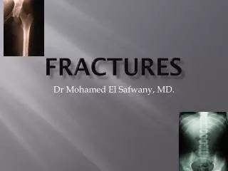
How would you describe these fractures? Displaced, shortened distal ulnar fracture
2nd metacarpal comminuted fracture 3rd/4th metacarpal oblique fractures with Minimal displacement and angulation, 5th Metacarpal fracture, nondisplaced
Proximal tibial fracture with displacement, shortening Proximal fibular fracture, simple?
Proximal tibial fracture, laterally displaced, angulated Proximal fibular fracture, laterally displaced, angulated
Specific Fractures • Galeazzi • Radial shaft • Radio-ulnar joint disruption • Monteggia • Mid-shaft ulna • Radial head disloc
Why are physeal injuries important? • Prognosis of physis injuries • Fractures of radius/ulna – 3-4% growth arrest (Cannata et al, 2003) • Fractures of femur – 40-50% growth arrest • Undulating surface of distal femoral physis
Anatomy • The bone growth process is from utero until the end of puberty • At birth, only a few epiphyses have begun to ossify, the rest are cartilaginous and are not visible on xray • As the child grows, other epiphyses begin to ossify and enlarge. • The epiphyseal plates (physes) are sites of cartilaginous proliferation/growth, and do not begin to ossify/close until puberty (earlier in females)
Anatomy • When skeletal injuries involve sites where ossification has not begun or is incomplete, xray may appear normal. May need MRI • Before closure of the growth plate in puberty, the growth plate is weaker than nearby ligaments • Injuries near the joints more likely result in physeal disruption/avulsion fractures than ligamentous tearing.
Evaluation of Fractures • History: mechanism of injury, location of pain, previous orthopedic history, past medical history, medications • If open fracture: tetanus history, where injury took place (dirty environment?) • PE: examine area of focus and the joint above and below • Examine for associated injuries
Fracture - Initial Assessment: • NPO • Immobilization • Pain medication • X-ray • Joint above and below injury • At least 2 views at 90° to each other
Evaluation of Fractures • Neurovascular status • Pulses (warmth, cap refill, extremity movement) • Sensation (compare to unaffected limb) • 6 “P’s” (pain, pulselessness, pallor, paralysis, paresthesias, painful passive mvmt): the presence of any of these, suggests neurovascular compromise
Evaluation of Fractures • Inspection: asymmetry, swelling, abrasions, ecchymoses, deformity • Joint exam: Active range of motion/passive • May be difficult to localize injury in children if unwitnessed fall • Children can self-splint injuries, making discomfort minimal
Evaluation of Fractures • Radiographs • AP/lateral views if obvious deformity, point tenderness, marked swelling/ecchymosis • Splint extremity prior to xrays • Views should include joints above and below • Include oblique view for toddler’s fx • May need comparison views (esp elbow)
Evaluation of Xrays • Follow the cortex to look for any discontinuity • Evaluate the growth plates and joints for displacement, disruption, or widening • May be difficult to differentiate between sprain and Salter I injury
Fractures suspicious for abuse • Metaphyseal (bucket-handle) • Rib (especially posterior) • Scapular • Spinous process • Sternal fractures • Epiphyseal separations • Vertebral body fxs and subluxations • Digital fxs
Fractures suspicious for abuse • Complex skull fxs • Multiple fractures (esp if bilateral or in various stages of healing) • Low specificity for abuse • subperiosteal new bone formation • Clavicular fxs • Fxs of long bone shafts • Linear skull fxs
ED Nondisplaced SH I Clavicular fractures Non-displaced UE fractures Incomplete/Non-displaced fractures of long bones of LE Nondisplaced hand/foot fractures Ortho Open fractures Unacceptably displaced fractures Neurovascular compromise Physeal/joint injuries Complete/displaced long bone fractures Pelvic & spinal fractures Joint sepsis ED vs Ortho Management
Management of Fractures ABC's!!!
Management of Fractures • Once vitals are stabilized, and patient examined completely for nonorthopedic injuries, must assess neurovascular status of effected limb. • If any of the 6 “P’s” present, suggests neurovascular compromise, get immediate orthopedic consult • Place extremity in longitudinal traction and align any gross deformities
Management of Fractures • If open fracture: culture exposed bone/soft tissue • Cover wound with sterile dressing • IV abx • Tetanus if applicable • Pain relief (splint extremity in a physiologic position, elevation, ice application) • Tylenol/ pain meds • XRAYS
Management of Fractures • Most nondisplaced extremity fractures can be treated with in situ immobilization with cast or splint • Displaced fxs or growth plate fxs: orthopedic consult • Reduce finger dislocations promptly: digital block, axial traction
Management of Fractures • Elbow/hip/knee dislocations are at risk neurovascular compromise, therefore require ortho consultation • Contusions/Sprains: RICE therapy, most injuries will resolve in 5-7 days • Severe sprains: commercial splint/Jones dressing (Webril/ace bandages) • Cast care
Clinical Scenario #1 • An 8 yo boy, complains of right upper chest pain after running into another player during football practice • Pain with right arm movement • Pain relieved with cradling the right arm with his left
Clinical Scenario #1 • Exam: TTP over right clavicular area • Pain with right shoulder abd/adduction • NV intact • No pain over humerus/radius/ulna/shoulder • What do you suspect? • Which xrays?
Clavicle Fracture: • Shaft • Fall on outstretched hand • Greenstick fractures • Callus formation • Sling or Figure of 8 brace • Lateral • Cortex remains intact • Risk of non-union • Medial (= sternoclavicular joint separation in adults) • Epiphyseal separation • Airway or vascular injury
Immobilization in a figure-of-eight dressing or a sling and swathe for 3 Weeks, followed by 3 weeks of restricted activity
Clinical Scenario #2 • A 6 yo girl presents with L elbow pain • Hx: fell from monkey bars at school, landing on L arm • Exam: L elbow TTP, pain with L elbow range of motion, CR<2, L hand warm, well perfused • NV intact
Clinical Scenario #2 Supracondylar fracture
Clinical Scenario #3 • 6 mo M, crying inconsolably for 4 hours at home today. • Mom says she is just learning how to roll over and thinks she twisted her leg while rolling over to her stomach • Afebrile, +URI symptoms, decreased energy • On exam, crying inconsolably, tachycardic, worsening cry with L leg passive movement • Point tenderness over L mid femur
Clinical Scenario #3 Femur fracture Needs spica cast, eval for possible abuse
Supracondylar Fracture • Fall on outstretched arm with hyper-extension of elbow • Posterior displacement of distal fragment commonly occurs • Complications • NV compromise • Deformity • Range of motion abnormalities
Supracondylar Fracture • Type I – nondisplaced • Type II – Angulated, posterior cortex intact • Type III – Disrupted posterior cortex w/ completely displaced distal fragment