Download
1 / 12
120 likes | 136 Vues
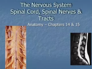
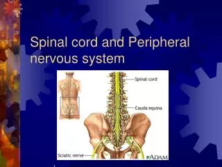
IV. Central Nervous System: Spinal Cord. A. Spinal Cord Location. 1. Enclosed within the vertebral column 2. Extends from the medulla oblongata to the region of T12 3. Below T12 is the cauda equina “horses tail” (a collection of spinal nerves)

E N D
A. Spinal Cord Location 1. Enclosed within the vertebral column 2. Extends from the medulla oblongata to the region of T12 3. Below T12 is the cauda equina “horses tail” (a collection of spinal nerves) • Enlargements occur in the cervical and lumbar regions Figure 7.18
Spina Bifida • At 21 days after conception (left drawing), folds of tissue on the back of a developing embryo are rapidly growing together (see lines) to form the neural tube. Just a day later (center drawing), the growth is almost complete. If the tissue fails to close completely (right drawing), development of the spine, muscle and skin in this region is affected and the baby will be born with spina bifida.
Spina Bifida Surgery • http://www.youtube.com/watch?feature=player_detailpage&list=FLw79wcj2687CXBNNaSAdOdQ&v=E080qJuHWdQ Uploaded by mayoclinic on Oct 30, 2008 Every year, thousands of babies are born with spina bifida. It's a birth defect where the spinal column does not close properly, exposing nerves to the environment.
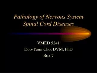
B. Spinal Cord Anatomy • Exterior white mater – conduction tracts Figure 7.19
B. Spinal Cord Anatomy • Internal gray matter - mostly cell bodies • Dorsal (posterior) horns- contain association neurons, or interneurons • Anterior (ventral) horns – contain cell bodies of motor neurons of somatic (voluntary) nervous system Figure 7.19
B. Spinal Cord Anatomy 1. Central canal filled with cerebrospinal fluid Figure 7.19
B. Spinal Cord Anatomy 2. Meninges cover the spinal cord to cushion and protect 3. Nerves leave at the level of each vertebrae • Dorsal root – sensory neurons • Associated with the dorsal root ganglia – collections of cell bodies outside the central nervous system • Ventral root – motor neurons 4. The dorsal and ventral roots fuse to form spinal nerves 5. 31 pairs of spinal nerves arise from the cord and exit from vertebral column
B. Spinal Cord Anatomy • 31 spinal nerves • 8 pairs cervical nerves • 12 pairs thoracic nerves • 5 pairs lumbar nerves • 5 pairs sacral nerves • 1 pair coccyx nerve