Download
1 / 69
700 likes | 988 Vues
The Menstrual Cycle & Pregnancy. Honors anatomy & physiology The reproductive system Part 3. Female Reproductive Cycle. 2 parts: Ovarian Cycle series of events in ovaries occurring during & after maturation of oocyte Uterine (Endometrial) Cycle concurrent with ovarian cycle

E N D
The Menstrual Cycle&Pregnancy Honors anatomy & physiology The reproductive system Part 3
Female Reproductive Cycle • 2 parts: • Ovarian Cycle • series of events in ovaries occurring during & after maturation of oocyte • Uterine (Endometrial) Cycle • concurrent with ovarian cycle • series of changes in endometrium to repare it for implantation of fertilized egg
Hormonal Regulation Hypothalamus secretes GnRH (gonadotropin-releasing hormone) that controls both the ovarian & uterine cycles GnRH stimulates release of FSH & LH from anterior pituitary
Effects on Ovaries FSH LH • initiates follicle growth • stimulates: • follicles to release E • granulosa cells to turn androgens E • stimulates : • further development of follicles • release of E from follicles • release of androgens from theca cells • @ midcycle LH surge triggers ovulation • corpus luteum E, P, relaxin, inhibin
Estrogens • 3 of 6 different estrogens are present in significant amts: • ß estradiol • Estrone • Estrial • most abundant in non-pregnant female • made from cholesterol in ovaries
Functions of Estrogens promote development &maintenance of female reproductive structures, 2◦ sex characteristics increase protein anabolism working synergistically with hGH lowers cholesterol (1 reason females <50 yo have lower risk of CAD) moderate levels inhibit release of GnRH, FSH, LH (negative feedback loop)
Progesterone secreted by corpus luteum with E prepares endometrium for implantation & breasts for lactation
Relaxin produced by corpus luteum inhibits contractions of myometrium during pregnancy, placenta secretes more relaxin…@ end of pregnancy it increases flexibility of pubic symphysis & may help dilate cx
Inhibin secreted by granulosa cells of growing follicles & by corpus luteum after ovulation action: inhibits secretion of FSH & to lesser extent LH
Phases of the Menstrual Cycle • Menstrual Phase • Preovulatory Phase • in ovaries called follicular phase • in uterus: proliferative phase • Ovulation • Postovulatory Phase • in ovaries called luteal phase • in uterus: secretory phase
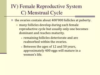
Menstrual Phase • aka menstruation, menses • ~1st 5 d of cycle (d 1 of flow = day 1 of cycle) • Ovaries: • FSH causes several primordial follicles to develop 1◦ follicles 2◦ follicles • Uterus: • declining E & P levels prostaglandins constriction of uterine spiral arterioles O deprived tissue death & shedding 50 – 150 mL of blood, tissue fluid, mucus, epithelial cells (entire stratum functionalis sloughs off)
Preovulatory Phase • time between end of menses ovulation • most variable length in time (reason cycles vary in length) • Ovaries: • follicles secrete E & inhibin d 16 • one 2◦ follicle in 1 of the 2 ovaries becomes the dominant follicle (secretes more E & inhibin decreases FSH) • dominant follicle (Graafian) enlarges until ovulation:~2cm
Preovulatory Phase (Proliferative) • Uterus: • E stimulates repair of endometrium producing new stratum functionalis • new endometrial glands form • endometrium thickens from ~4 to 10 mm
Ovulation d 14 in 28 d cycle 2◦oocyte surrounded by zonapellucida & corona radiata higher levels of E @ end of preovulatory phase have + effect on cells that secrete LH & GnRH: their increase leads to ovulation OTC test for LH surge used to predict ovulation
Mittleschmerz pain noted @ time of ovulation caused by small amt of blood that leaks into pelvic cavity from ruptured follicle
Postovulatory Phase (Luteal) • time between ovulation & onset of next menses • Most constant part of cycle = 14 days (d15 – 28) • Ovary: • under influence of LH, granulosa cells transformed corpus luteumcellswhich secrete E, P, relaxin, inhibin • if oocyte not fertilized: corpus luteum lasts 2 wks corpus albicans • + fertilization: corpus luteum persists until “rescued” by hCG (human chorionic gonadotropin) which is made by chorion of embryo @ ~ 8d after fertilization
Postovulatory Phase (Secretory) • Uterus: • P & E made by corpus luteum promote: growth & coiling of endometrial glands • vascularization of superficial endometrium • thickening of endometrium (12 – 18 mm) • these changes peak 1 wk after ovulation • if no fertilization levels of P & E decline which eventually causes menstruation
Homeostatic Imbalances • Dysmenorrhea • painful menses • Amenorrhea • absence of menses • #1 reason = pregnancy • Endometriosis • disorder in which endometrium grows outside uterus • +/- painful • can cause infertility due to scarring
Embryonic Period • Fertilization: • Nuclei from sperm merges with nuclei from 2◦oocyte forming a diploid nucleus • Fallopian tube normal site of fertilization w/in 12 -24 hrs after ovulation • sperm can remain viable up to 48 hrs after deposition in vagina
Fertilization • sperm must penetrate 2 layers: • corona radiata • granulosa cells that surround 2◦oocyte • zonapellucida • clear glycoprotein layer between corona radiata & oocyte’s plasma membrane
Fertilization ZP3: 1 of glycoproteins acts as sperm receptor acrosomal reaction: occurs when ZP3 binds to specific membrane protein on sperm head plasma membrane release of contents of acrosome
Acrosomal Reaction acrosomal enzymes digest a path thru zonapellucida lashing flagella of sperm pushes it forward several sperm bind to ZP3 molecules but only 1st sperm to penetrate zonapellucida & reach plasma membrane of oocyte “wins” once diploid nucleus formed its called a zygote
Cleavage of the Zygote rapid M phase but no growth 1st division begins ~24 hrs after fertilization taking 6 hrs to complete, following divisions take less time ~2 d after fertilization = 4 cells ~ 3 d after = 16 cells cells get progressively smaller, & are called blastomeres morula (mulberry) solid sphere of cells, still surrounded by zonapellucida & still about size of original zygote
Blastocyst Formation ~ end of 4th d: # of cells in morula increase as it is still moving thru fallopian tube enter uterine cavity on d 4-5 glycoprotein secretions fromendometrial glands enter morula providing nourishment @ ~ 32 cell size, fluid enters morula & collects between blastomeres forming fluid-filled cavity = blastocyst cavity (up to ~100 cells) & now called a blastocyst or blastula (still ~ same size as original zygote)
Blastocyst Forms Layers • 2 distinct structures form: • Inner cell mass • eventually becomes embryo • Trophoblast • ultimately forms fetal portion of placenta • 5th d: blastocyst digests hole thru zonapellucida & squeezes out
Implantation attachment of blastocyst to endometrium after implantation endometrium becomes modified & is called decidua
Trophoblast develops into 2 layers (both part of chorion): Syncytiotrophoblast Cytotrophoblast
hCG is Produced hCG: human chorionic gonadotropin Produced by trophoblast starting on d 6 hCG causes endometrium to grow & proliferate hCGprevents the menstrual cycle from occuring reason female misses her menses when she is pregnant
Inner Cell Mass differentiates into a bilaminar disc Hypoblast Epiblast
Amnion thin protective membrane develops from cytotrophoblast
Gastrulation @ end of cleavage stage, cells making up the blastula move surface proteins help cells recognize each other & help sort cells 3 layers of gastrula formed: called the 3 germ layers Endoderm Mesoderm Ectoderm
Extraembryonic Membranes develop from the germ layers but are NOT part of the embryo (lost at birth) lie outside embryo & provide protection & nourishment 4 components: chorion amnion allantois yolk sac
Placentation formation of the placenta site of exchange of nutrients & wastes between the mother & fetus also functions as protective barrier & produces several hormones to maintain pregnancy (hCG)