Download
1 / 19
230 likes | 514 Vues
Brainstem. Components of Brainstem : Midbrain , Pons & Medulla. Dr Taha Sadig Ahmed. Midbrain The midbrain is divided into three parts: 1- Tectum It includes:

E N D
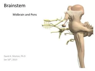
Brainstem Components of Brainstem : Midbrain , Pons & Medulla Dr TahaSadig Ahmed
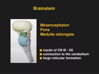
Midbrain • The midbrain is divided into three parts: 1- Tectum • It includes: • A- The superior colliculus. It is involved in vision and sends its superior brachium to the lateral geniculate body of the thalamus • B- The inferior colliculus, is involved in hearing & sends its inferior brachium to the medial geniculate body of the thalamus. • The Cerebral Aqueduct runs through the midbrain, beneath the colloculi. • 2- Tegmentum • Liesventral to the cerebral aqueduct. Several nuclei, tracts and the reticular formation is contained here. • 3- The ventral side of the midbrain • is comprised of paired cerebral peduncles. These transmit axons of UMN.
Midbrain internal structures • Periaqueductal Gray: Around the cerebral aqueduct, contains neurons involved in analgesia & pain modulation/desensitization . • Occulomotor nerve (CN III) nucleus , which controls movements of some eye muscles . • Trochlear nerve (CN IV) nucleus which also controls movements of some eye muscles . • Red Nucleus: gives out Sends Rubrospinal tract which is inhibitory to spinal Gamma Efferents neurons ( & stretch reflex /muscle tone ) • SubstantiaNigra: Collection of neurons in the ventral portion of the midbrain . They secrete Dopamine (which is involved in motor function ). Degeneration of SubstantiaNigra is associated with Parkinson’s disease .
Central Tegmental tract: Directly anterior to the floor of the 4th ventricle, this is a pathway by which many tracts project up to the cortex and down to the spinal cord • Reticular formation: • A large area that contains ascending & descending fibers and is involved in involved in • Pain modulation/desensitization • Consciousness and arousal • contains the Locus Ceruleus, which is involved in autonomic reflexes
Ventral view of the pons • •Between the basal pons, cranial nerve 6 (abducens), • 7 (facial) and • 8 (vestibulocochlear) emerge • (medial to lateral). • •At the level of the midpons, the large trigeminal nerve, CN V, emerges.
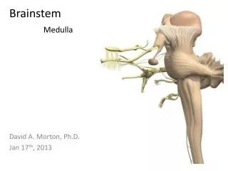
Ventral view of the medulla • The most medial part of the medulla is the anterior median fissure. • Moving laterally on each side are the pyramids. • The pyramids contain the fibers of the corticospinal (pyramidal) tract as they head inferiorly to synapse on lower motor neuronal cell bodies within the ventral horn of the spinal cord. • The anterolateralsulcus is lateral to the pyramids.
Emerging from the anterolateralsulci are the hypoglossal nerve (CN XII) rootlets. • Lateral to these rootlets and the anterolateralsulci are the olives. • The olives are swellings in the medulla containing inferior olivary nuclei • Lateral (and dorsal) to the olives are the rootlets for cranial nerves IX and X (Glossopharyngeal and Vagus, respectively
Dorsal view of the medulla • •The most medial part of the medulla is the posterior median fissure. • Moving laterally on each side is the Fasciculus Gracilis, and lateral to that is the Fasciculus Cuneatus. • Superior to each of these, are the gracile and cuneate tubercles, respectively. Underlying these are their respective nuclei • In the midline is the VagalTrigone and superior to that is the Hypoglossal Trigone. Underlying each of these are motor nuclei for the respective cranial nerves
Functions of the brain stem • Though small, brain stem is an extremely important part of the brain: • 1. It has conduct functions. • 2. Provides the origin of the cranial nerves (CN III-XII). • 3. Conjugate eye movement. • 4. Integrative functions.
1. Conduct Functions of the Brainstem • All information related from the body to the cerebrum and cerebellum and vice versa, must traverse the brain stem • A- Ascending ( sensory ) tracts coming from the body to the brain include • (1) Spinothalamic tract for pain and temperature sensation. • (2) Dorsal column tracts , fasciculus Gracilis, and fasciculus Cuneatus for touch , & Proprioceptive and Pressure sensation.
Ventral Surface Dorsal Surface
B- Descending (MOTOR) tracts are • The Corticospinal ( Pyramidal ) tract : • It is an Upper Motor Neuron ( UMN) that originates in the cerebral cortex • Its fibers runs through the CrusCerebri, the basal part of the Pons and the Medullary Pyramids; • 70-90 % of its fibers cross in the pyramidal decussation to form the lateral corticospinal tract, destined to synapse on lower motor neurons in the ventral horn of the spinal cord • other UMNs that originate in the Brainstem from (1) vestibular nucleus , (2) Red nucleus , and (3) Reticular nuclei, also descend and synapse in the spinal cord
2. The brain stem provides the main motor and sensory innervation to the face and neck via the cranial nerves (CN III-XII). The fibers of cranial nerve nuclei except for olfactory & optic nerve either originating from, or terminating in, the cranial nerve nuclei in brain stem.
Origin & functions of the cranial nerves • From Midbrain • CN III (Oculomotor) • CN IV (Trochlear) • Both moves eyes; CN III contsricts the pupils , & accomodates • From Pons • •CN V (Trigeminal) : Chewing ( mastication ) and sensation from front of the head. • •CN VI (Abducens) : Some eye movement • •CN VII (Facial): (1) controls facial muscles ( facial expressdion ) (2) Taste • (3) Salivation (4) Tears • •CN VIII (Acoustic , Vestibulo cochlear): Hearing & Balance
From Medulla • CN IX (Glossopharyngeal): Tastes, salivates, swallows, monitors carotid body and sinus. • CN X (Vagus): Tastes, swallows, lifts palate, talks, communication to and from thoraco-abdominal viscera. • CN XI (Accessory): Turns head, lifts shoulder. • CN XII (Hypoglossal): Moves tongue • Classification of Cranial Nerves According to Function • Sensory • First Cr n ( Olfactory) , 2nd ( Optic ) , 8thVestibulocochlear • •Motor • 3rd (Oculomotor) , 4th ( Trochlear) , 6th ( Abducens) , 11th ( Accessory ) , 12th ( Hypoglossal) • •Mixed • 5th ( Trigeminal ), 7th ( Facial),9th (b Glossopharyngeal) ,10th Vagus
Integrative Functions of Brainstem • (1) It controls consciousness & sleep cycle (alertness and arousal) through reticular formation (RAS). • (2) It has got center for cardiovascular, respiratory & autonomic regulation . • (3) It has centers for Brainstem Reflexes , such as cough reflex , gag reflex , swallowing , and vomiting ; + visual & auditory orientation reflexes (required for head movements. through Superior & Inferior Colliculi ) • (4) Contributes to maintenance of body balance through the vestibular nuclei • (5) Plays role in motor control • (i) SubstantiaNigra( which is a part of the basal ganglia ) is involved in control of movement. • (ii) Midbrain also contain Red nucleus which regulate the motor activity through cerebellum
Control of Conjugate Eye Movements • Conjugate eye movement refers to motor coordination of the eyes that allows for bilateral fixation on a single object. • Several centers in the brainstem are involved. • Horizontal conjugate gaze is controlled by • (1) the nuclei of 3rd and 6th Cranial nerves , • (2) the Paramedian Pontine Reticular Formation, and • (3) the nucleus prepositushypoglossi-medial vestibular nucleus. • Vertical conjugate gaze is controlled by the nuclei of • 3rd and 4th Cranial nerves
Pain sensitivity control: Periaqueductal Grey Matter of mesencephalon ( midbrain ) is an area which is rich in endogenous opioid and is important in modulation of painful stimuli. • Layers of Brainstem • Ventral layer of brainstem is motor in function. • Middle layer is sensory in function & contains medial lemniscus which conveys sensory information from dorsal column.
Brain Stem Function Tests (A) To test reticular formation (1) Alertness, Consciousness & Sleep. (2) Corticospinal tract (3) Motor power, reflexes (4) Pain response Facial grimacing on firm pressure over the supra orbital ridge. (B)To test respiratory center look for the normal pattern of respiration (C)To test cardiovascular center : Look for normal circulatory function (D)To test brainstem reflexes: • Pupilary and corneal reflexes. • Vestibulo-ocular reflex: Injection of iced water into the ear will produce eyes movement. • Oculo-cephalic reflex: Eyes will be fixed when head is moved in one or another directions. • Gag reflex. Cough