Download
1 / 51
530 likes | 594 Vues
Slides prepared and compiled by highly experienced ENT teacher, Dr. Krishna Koirala from Nepal, for teaching undergraduate and postgraduate ENT students in the field of otorhinolaryngology. <br>A clear and concise explanation of the basic concepts in the subject matter concerned. <br>He is the Head of department with a sound knowledge in the field of ENT to teach both undergraduate and postgraduate ENT students<br>

E N D
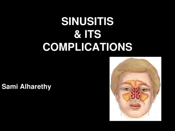
Complications of Sinusitis Dr. Krishna Koirala 2018-08-13
Definition • Progress of infection beyond the muco-periosteal lining of paranasal sinuses to involve the bone and neighboring structures (orbit, intra-cranial cavity, dentition) • Compromise in function of any part of body due to sinusitis
Etiology • Weak immune response of host • Young children and immuno -compromised adults • Inadequate / inefficient treatment • Infection by highly virulent organisms • Abnormalities of muco- cilliary clearance • Persistent allergy and blockade of sinus ostia
Routes of infection • Via thin bones eg. lamina papyracea • Through natural suture lines • Through natural canal: infra-orbital canal • Retrograde thrombophlebitis: diploic vein of Breschet • Closely related roots of upper 2nd premolar & 1st molar teeth • Periarteriolar spaces of Virchow Robin
Classification • Acute • Local • Orbital • Intracranial • Bony • Dental • Distant • Toxic shock syndrome • Chronic • Mucocele • Pyocele • Associated diseases (?) • Otitis media • Adeno -tonsillitis • Bronchiectasis
Orbital Complications ( Chandler et al 1970) 1. Pre-septal cellulitis 2. Orbital cellulitis without abscess 3. Orbital cellulitis with extra/ sub-periosteal abscess 4. Orbital cellulitis with intra-periosteal abscess 5. Cavernous sinus thrombosis
Intracranial Complications 1. Meningitis 2. Encephalitis 3. Extra-dural abscess 4. Sub-dural abscess 5. Intra-cerebral abscess 6. Cavernous sinus thrombosis 7. Sagittal sinus thrombosis
Bony • Osteitis • Osteomyelitis (Pott’s puffy tumour) • Dental • Dental abscess • Oro-antral fistula
Orbital complications • Commonest complication of sinusitis • Young people at high risk: 85% < 20 yrs age • Ethmoid sinus most commonly implicated Frontal Sphenoid Maxillary • Left orbit more commonly involved (?)
Pre-septal cellulitis • Inflammation external to orbital septum • Edema of eyelids: • Upper lid : frontal sinusitis • Lower lid : maxillary sinusitis • Both lids : ethmoid sinusitis • No tenderness , visual loss , limitation of extra-ocular movement
Orbital Cellulitis without abscess • Inflammation of adipose tissue deep to peri-orbital septum without suppuration • Diffuse peri -orbital edema with erythema • Mild proptosis • No restriction of extra-ocular movement • No change in vision
Extra-periosteal abscess • Most common form of orbital cellulitis • Localized extra-periosteal pus collection • Mild proptosis, restriction of extra-ocular movement , vision loss • Color vision affected first • Red = brown • Blue = black
Orbital cellulitis with Intra-periosteal abscess • Mild chemosis (edema of conjunctiva) • Proptosis: severe, asymmetric, quadrantic • Frontal sinusitis : down + forward + lateral • Ethmoid sinusitis :forward + lateral • Maxillary sinusitis : up + forward • Concurrent and complete ophthalmoplegia • Visual loss due to optic neuropathy (up to 13% of cases)
Cavernous Sinus Thrombosis • Rapid onset, hectic fever • Bilateral orbital pain + severe chemosis • Bilateral absent pupillary reflex • Bilateral symmetrical axial proptosis • Sequential ophthalmoplegia (VI III IV) • Papilledema + loss of vision • Painful paresthesia of V1, V2
Evaluation of orbital complication • Ophthalmology consultation • Look for edema of eyelids, displacement of eyeball (proptosis),restriction of ocular movement • Visual acuity and color vision examination • Fundoscopy for papilledema • CT scan PNS (including orbit): coronal and axial cuts
Medical Treatment • Broad spectrum, high dose IV antibiotics • Ceftriaxone + Metronidazole+ Amikacin • NSAIDs • Topical / oral nasal decongestants • Mucolytics:Bromhexine, Ambroxol, Guaphanesin • Nasal saline irrigation
Surgical Treatment • For sinusitis • Frontal sinus trephination • External fronto-ethmoidectomy (Lynch Howarth) • Functional Endoscopic Sinus Surgery ( FESS) • For orbital complications • Sub-periosteal abscess drainage • Orbital decompression
Introduction • 2nd most common complication of sinusitis • Most common in adolescents & young adults (diploic venous system at peak vascularity) • Frontal sinus most commonly implicated Ethmoid Sphenoid Maxillary • Commonest route of spread : Retrograde thrombophlebitis via Diploic vein of Breschet
Clinical Features • Fever • Deep-seated headache • Nausea & projectile vomiting • Neck stiffness • Seizures • Altered sensorium & mood changes • Late: bradycardia / hypotension / stupor
Investigations and Medical Treatment • Neurosurgery consultation • CT scan PNS + brain with contrast • MRI with contrast: investigation of choice • High dose broad spectrum I.V. antibiotics: Ceftriaxone & Metronidazole for 4-6 week • Steroids : controversial
Surgical treatment for abscess • For sinuses: • Frontal trephination • External fronto-ethmoidectomy (Lynch Howarth) • Functional Endoscopic Sinus Surgery • For intra-cranial complication: by Neurosurgeon • Burr hole drainage for small abscess • Craniotomy for large brain abscess
Introduction • Definition: epithelium lined, mucus filled sac filling the paranasal sinus that is capable of expansion • Incidence: • Frontal : 65 % • Ethmoid : 25 % • Maxillary : 10 % • Sphenoid : rare
Etiology • Chronic obstruction of sinus ostium with retention of normal sinus mucus within sinus cavity • Mucous retention cyst : Develops from obstruction of ducts of sero mucinous glands within sinus mucosa
Clinical Features • Cystic, non-tender swelling above inner canthus with egg-shell crackling sensation on palpation • Proptosis: • Frontal :downward + forward + lateral • Ethmoid : forward + lateral • Maxillary : up + forward • Diplopia & restricted eyeball movement • Frontal headache, retro-orbital or facial pain
Investigations • X-ray PNS OM view: expanded frontal sinus, loss of scalloped margins, translucency, depression or erosion of supra-orbital ridge • CT scan:homogenous smooth walled mass expanding the sinus with thinning of bone • Ring enhancement on contrast: pyocoele
Treatment 1. Antibiotics and nasal decongestants 2. External fronto-ethmoidectomy by Lynch – Howarth’s approach 3. Endoscopic fronto-ethmoidectomy 4. Endoscopic decompression (marsupialization) 5. Osteoplastic flap repair
Drainage + Marsupialization Post-op CT scan (coronal)
Pott’s puffy tumour • Frontal sinus osteomyelitis (Percival Pott, 1760) • Fluctuant swelling over forehead anteriorly • May spread posteriorly leading to subdural abscess • Treatment • Six week course of broad spectrum antibiotics • Drainage of pus & debridement of bone • Obliteration of frontal sinus by osteoplastic flap technique
Oro-antral fistula • Fistulous tract communicating between oral cavity and maxillary antrum • Treatment : closure by • Buccal mucosal advancement flap • Palatal flap • Buccal fat pad flap