Download
1 / 54
570 likes | 918 Vues
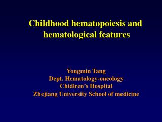
Lecture 3. Hematopoiesis : The process to produce immune cells. CORE. 6-b. Hematopoiesis. Stem cells and cytokines Hematopoiesis in bone marrow is regulated by some cytokines such as stem cell factor, IL-1, IL-3, IL-6, IL-7, GM-CSF, EPO, G-CSF and M-CSF. Figure 1-10.

E N D
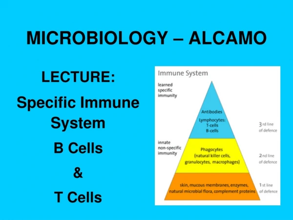
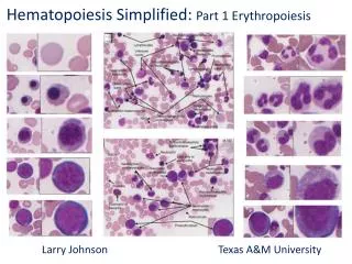
Lecture 3. Hematopoiesis: The process to produce immune cells
CORE 6-b. Hematopoiesis • Stem cells and cytokines Hematopoiesis in bone marrow is regulated by some cytokines such as stem cell factor, IL-1, IL-3, IL-6, IL-7, GM-CSF, EPO, G-CSF and M-CSF
Figure 1-10 Hematopoiesis occurs in the adult bone marrow
Hematopoiesis generates immune cells Stem cells: 1. Self renewal 2. Totipotency They are in bone marrow after fetal development. They make all myeloid and lymphoid immune cells and RBCs T cell progenitors migrate to thymus and generate T cells B cell progenitors reside in bone marrow to make naïve B cells
Myeloid vs. Lymphoid cells Stem cells Myeloid cells Lymphoid cells T cells: T cell antigen receptor B cells: B cell antigen receptor NK cells: no antigen-specific receptor
Neutrophils: Phagocytes • Origin and maturation: Bone marrow • Antigen receptors: No • Function: Phagocytosis and killing of microorganisms • Where: in blood circulation • Sites of function: infection sites • Short life span Induced by the cytokines IL-3, GM-CSF and G-CSF
Monocytes: Macrophage precursors • Origin : bone marrow • Antigen receptors: No • Function: to become macrophages • Present in blood circulation IL-3, GM-CSF and M-CSF induce monocytes
Eosinophils: worm (parasites) killers • Origin : bone marrow • Antigen receptors: No • Function: killing of antibody-coated parasites through release of killing mix (cytotoxic granules) • Effector machinery:cytotoxic granules, lipid mediators, cytokines and chemokines IL-5 induces eosinophils
Mast cells: parasite killers • Origin : bone marrow • Antigen receptors: No • Function: to kill parasites • Sensor: IgE receptor • Effector machinery:cytotoxic granules, lipid mediators, cytokines and chemokines • Found in connective tissues Stem cell factor (SCF) induces mast cells
Basophils: relatives of mast cells and eosinophils • Origin : bone marrow • Antigen receptors: No • Function: important effector cells in allergic disorders and immune responses to parasites • Sensor: IgE receptor • Effector machinery:cytotoxic granules, lipid mediators, cytokines and chemokines
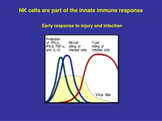
NK cells: natural killers • Origin : Bone marrow and thymus • Antigen receptors: No • Function: Kill tumor and virus-infected cells • Effector machinery (=weapons): perforins and granzymes • Activating or inhibitory receptors
T lymphocytes: master regulators of the immune system • Origin: Bone marrow • Maturation: Thymus • Differentiation to effector cells in secondary lymphoid tissues (lymph nodes, spleen, Peyer’s patch, and tonsils) • Antigen receptors: Yes • Function: regulates humoral and cell-mediated immune responses • Mechanisms: cytokines, cell surface molecules, cytotoxic granules.
B lymphocytes: antibody producers • Origin and maturation: Bone marrow • Differentiation to plasma B cells in secondary lymphoid tissues (lymph nodes, spleen, Peyer’s patch, and tonsils) • Antigen receptors: B cell receptor (cell surface immunoglobulins) • Function: Production of antibodies (IgM, IgE, IgA, and IgG) • Regulated by T cells
B lymphocytes Antigens+ T cell help
Figure 1-12 Circulating blood cells
Hematopoietic stem cell therapy & cytokine therapy Bone marrow transplantation into immunodeficient patients or cancer patients after radiation/chemotherapy Recombinant G-CSF and GM-CSF for neutropenia (neutrophils and monocytes) G-CSF: Neupogen™ and Neulasta™ GM-CSF: Leukine™ Help prevent infection by boosting leukocyte production. Recombinant erythropoietin (EPO) for anemia (red blood cells) EPOGEN®
Lecture objectives • Big questions? How TCR recognizes antigens? What are the autoimmune diseases that are associated with particular types of MHC genotypes? Why MHC molecules are the major antigens responsible for transplantation rejections? To know: • Names of human MHC I and II genes. • How antigens are processed for presentation on MHC I and II? • Endogenous antigens and exogenous antigens? • MHC class I and II molecules present antigens of different origins. How? • MHC polymorphism • How many different MHCs a person can express? Why? • Structures of MHC I and II • HLA typing
A big picture:How do T cells recognize antigens? MHC molecule TCR
CORE • Major Histocompatibility Complex (MHC; Human Leukocyte Antigens [HLA]) • a. Class I , II and III MHC genetic loci (short arm of chromosome 6) • Major Class I genes: HLA-A, B, C • Minor Class I-like genes: HLA-E, F, G, H, J, X • Major Class II genes: HLA-D region • DP (A1, A2, B1, B2), DQ (A1, A2, B1, B2, B3), DR (A, B1, B2, B3) • Major Class III genes: Diverse (non-antigen presenting functions)
Genes in yellow: Functional MHC II genes Genes in dark gray: pseudogenes (not expressed, so not functional)
CORE c. Structure of Class I MHC proteins (1) a1, a2 and a3 domains of heavy chain (a1 & a2 form peptide binding site [groove]; amino acid differences account for polymorphism and antigen specificity) (2) b2 - Microglobulin (invariant but essential)
CORE • Structure of Class II MHC proteins • (1) Composed of one a and one b chain • (2) a1 and b1 domains comprise the peptide binding site (groove). Again, amino acid differences account for polymorphism and antigen specificity. a2 and b2 domains are constant.
Figure 3-2 T CELL RECEPTOR
Figure 3-15 The peptide-binding groove of MHC molecules
CORE • Inheritance: • Definition of haplotype, • Example of inheritance pattern, • Pseudogenes • Cell surface expression (which cell types express Class I, Class II). • Polymorphic nature of the MHC proteins (allotypes and gene polymorphism).
CD8 T or NK cells NK cells Remains intracellular NK cells MHC CLASS I molecules form ligands to activate CD8+ cells and inhibit NK cells
Polymorphism: presence of multiple alternative forms (alleles) of a gene. Help peptide loading Present antigen peptides to CD4+ T cells Polymorphism allows the population can handle a variety of pathogens.
Genetics of MHC gene expression:both alleles are expressed (co-dominant) • In any mating, four possible combinations of haplotypes can be found in the offspring; thus siblings are also likely to differ in the MHC allele they express. • Halplotype: The particular combination of MHC alleles found on a given chromosome 6.
Present Ag to CD8 T cells Present Ag to CD4 T cells
Figure 3-29 Each MHC isoform binds a characteristic set of peptides-Anchor residues in peptides are important for binding to MHC-Not all residues are important Degenerate binding allows each MHC molecule handles many different peptides.
Figure 3-26 Tom Jane Martin John
MHC: things to remember • MHC molecules in humans is also called HLA (human leukocyte antigen) • Class I and II loci. • HLA-DR alpha chain is monomorphic • HLA-DRB1 is most polymorphic in MHC II genes • HLA-DRB1 is always present in any individual • HLA-DRB3/4/5 is present in some but not all people. • A heterozygote person (most people) expresses two haplotypes. • A person can express 3-6 class I and 3-8 class II isoforms. • 2406 possible class II isoforms in the human population. • 753 MHC I isoforms in the human population. • [MHC isoforms] [presentable antigen peptides]
Figure 3-22 Different cell distribution of MHC class I and II • Almost all cells express MHC I for comprehensive surveillance by CD8 T cells • Only some cells express high levels of MHC II and MHC I • These are B cells, macrophages, dendritic cells and thymic epithelial cells. • B cells, macrophages and dendritic cells are called professional antigen- presentingcells (APC). • IFN-g increases the expression of MHC II in APC and induces the expression in non-APC cells at sites of infection
Processing and presentation of endogenous antigen via the MHC class I pathway (endogenous pathway): • Cytoplasmic proteins (e.g. viral proteins) are ubiquitinated, hydrolyzed to peptide fragments in the proteasome, and the peptides are transported into the ER via TAP. • 2. MHC I proteins are synthesized and assembled in ER and associated with TAP with the help from calnexin chaperone. • 3. MHC I proteins bind peptides, vesicles fuse with plasma membrane, and MHC I/peptide complexes are expressed on cell surface and presented to CD8 cells.
Processing and presentation of exogenous antigen via the MHC class II pathway (exogenous pathway): • Professional antigen processing cells internalize antigens. • Antigens are internalized into endosome & degraded into peptide fragments. • MHC class II proteins are synthesized in ER and the peptide binding site is protected by Ii (invariant chaperone; CLIP region of Ii protectsthe site). • Exocytic vesicles (from Golgi) containing MHC II proteins fuse with endosome containing peptides. • Ii is degraded, and CLIP is removed by HLA-DM protein. • MHC II proteins bind peptides, vesicles fuse with plasma membrane, and MHC II/peptides are expressed on cell surface and presented to CD4 cells.
Two different types of antigens:Extracellular for MHC II and intracellular for MHC IThey are processed differently.
Figure 3-9 Antigen processing is required to present antigen peptides to TCR.TCRs bind short antigen peptides but not whole antigen proteins
Figure 3-17 Transport of Cytosolic Peptides into ERRole of transporter associated with antigen processing, TAP Bare lymphocyte syndrome: No TAP expression no MHC I expression no CD8 T cells defective cytotoxic activity against virus-infected cells chronic respiratory infection TAP=TAP1+TAP2 Proteasome: a barrel shaped protein complex composed of 28 subunits 8 or more AA-long polypeptides are transported MHC I cannot leave ER without loaded peptides
Assembly of antigen peptide/MHC class I complex.Molecular chaperons (calnexin, calreticulin and tapasin)aid the folding of MHC I and loading of peptides Figure 3-18 In the absence of infection, self peptides are presented on MHC, but do not activate T cells. A HSV protein inhibits TAP function, and an Adeno virus protein inhibits MHC I expression.
Figure 3-20 Assembly of antigen peptide/MHC class II complex-Extracellular microorganisms are taken up by macrophages via phagocytosis and by B cells via cell surface Ig-mediated endocytosis-MHC II molecules bind peptides in the fused vesicles, not in ER-Invariant chain, CLIP and HLA-DM guide the peptide loading-After losing CLIP, MHC II must bind peptides or gets degraded.-Certain pathogens (e.g. mycobacteria), when engulfed, prevent the fusion of phagosomes and lysosomes, and persist in phagosomes.
Figure 3-12 MHC class I molecules present antigens to CD8+ T cells, and MHC class II molecules present antigensto CD4+ T cells:
Figure 3-30 T cell receptor recognition of antigens is MHC-restricted
TCR recognition of antigens induces T cell activation, functional maturation, and killing/activation of target cells Figure 3-11 The T cells cytokines are produced only when T cells are engaged with APC