Download
1 / 115
1.15k likes | 1.17k Vues
Radiographic Contrast. RTEC - A 2011 SUBJECT & FILM CONTRAST CONTRAST MEDIA. 2 types of Radiographic “Contrast”. Subject contrast patient Film contrast Inherent in equipment The BLACKS & WHITES ON THE FILM / IMAGE. “Subject” Contrast. Subject Contrast.

E N D
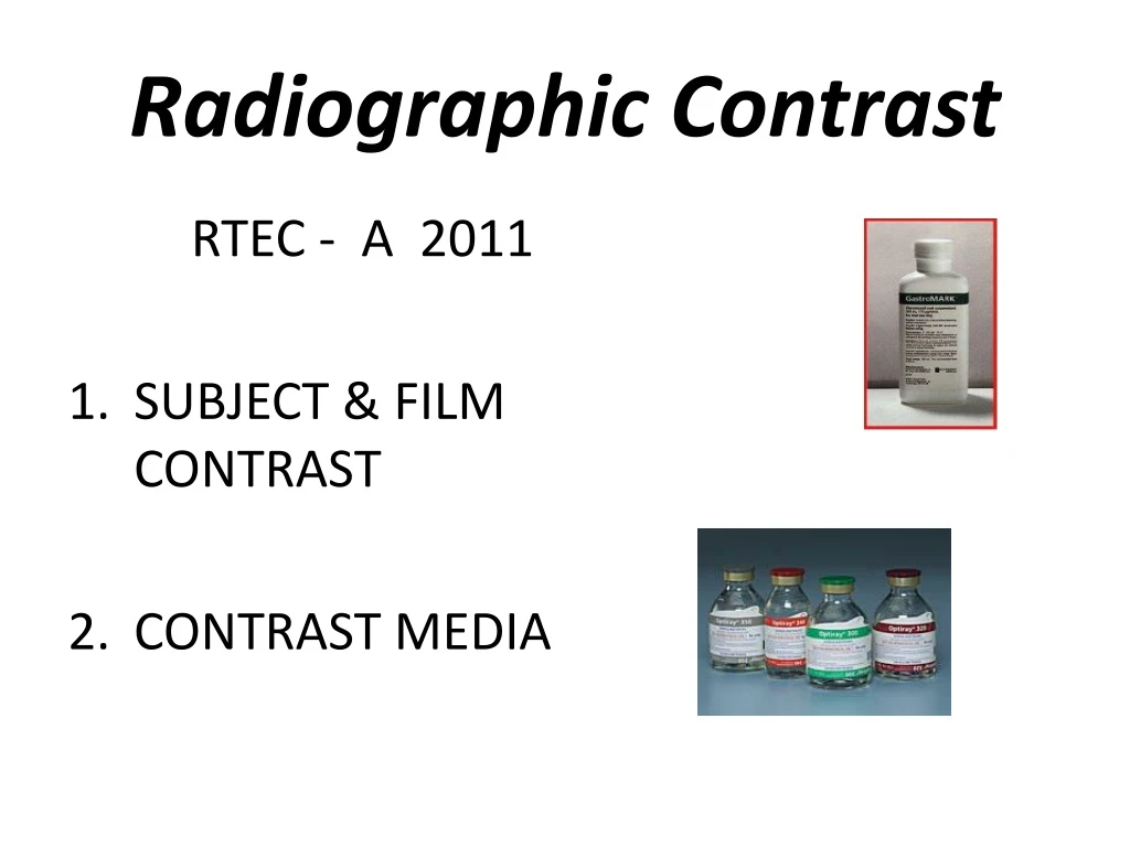
Radiographic Contrast RTEC - A 2011 • SUBJECT & FILM CONTRAST • CONTRAST MEDIA
2 types of Radiographic “Contrast” • Subject contrast • patient • Film contrast • Inherent in equipment • The BLACKS & WHITES ON THE FILM / IMAGE
Subject Contrast • Range of differences in the intensity of the x-ray beam • After it has been attenuated by the subject.
SUBJECT CONTRASTRadiographic object - influenced by • Atomic Number of object • Density of object • Thickness of object • 5 materials seen on a radiograph, • Gas/air, fat, soft tissue (muscle/organs), • bone and metals
Atomic Number • Fat = 6.46 • Water = 7.51 • Muscle = 7.64 • Bone = 12.31
TissueSubject Contrast • Atomic # of object • Density of object • Thickness of object • Higher atomic # = more attenuation • Denser = more attenuation • Thicker = more attenuation
PATHOLOGY • Pleural Effusion • Excessive fluid in lung • More dense than air
Pneumothorax • Lung collapses • No tissue in space • Easy to penetrate with x-ray photons
Radiographic Contrast influenced by: • Radiation Quality (KVP) • Film attributes • Radiographic object (Patient)
What is good contrast ? • High contrast (black and white) • Low contrast (more shades of gray)
RADIOGRAPHIC IMAGE Radiation Quality = kVp • High kVp ↑ 80 • Low contrast • Lots shades of gray • Long Scale • Little differences in adjacent structures • Low kVp ↓ 70 • High contrast • Black and White • Short Scale • Great differences in adjacent structures
Contrast changes with the use of a grid Less scatter radiation – shorter scale = “better contrast” With Grid No Grid
QUALITY – KVP • A visible change in contrast will not be seen until kVp is changed 4-12 % • kVp level change change in kVp • 30-50 kVp 4-5 % 1-3 kVp • 50-90 kVp 8-9 % 4-8 kVp • 90-130 kVp 10-12 % 9-16 kVp
Scenario • Low subject contrast in the area of interest. • You want to see the difference between muscle & fat & organs? • What can be done to attain medical information and define organ structure and function? • _____________________________________
Scenario • Low subject contrast in the area of interest. • You want to see the difference between muscle & fat & organs? • What can be done to attain medical information and define organ structure and function? • USE CONTRAST MEDIA
Purpose of Contrast Media • To enhance subject contrast or render high subject contrast • In a tissue that normally has low subject contrast. • Creates bigger differences in atomic number (z #’s)
Categories of Contrast Media Negative contrast • (AIR OR CO2) • Radiolucent • Low atomic # material • Black on film Positive contrast • (all others) • Radiopaque • High atomic # material • White on film
RADIOLUCENT - dark on image • AIR, CO2 • RADIOPAQUE - white on image • BARIUM • IODINE
AIR / CO2 Naturally seen in the LUNGS STOMACH (gas in intestines) Negative Contrast
2 BASIC TYPES OF ‘”Positive” CONTRAST MEDIA BARIUM Z# 56 KVP 90 – 120* • NON WATER SOLUABLE • GI TRACT ONLY INGESTED OR RECTALLY IODINE Z# 53 KVP BELOW 90* USUALLY 70 – 80 KvP • WATER SOLUABLE • POWDER • LIQUID • INTRAVENOUS OR • GI TRACT • OIL BASED • DUCTS /ORGANS
Positive Contrast Material INGESTED /INSTILLED • (ORALLY OR RECTALLY) • BARUIM • IODINES • GASTROGRAFIN • HYPAQUE POWDER INJECTED • IV – INTO BLOOD VESSELLS • Organs and ducts • IODINES • IONIC OR NON-IONIC • VESSELLS & ORGANS • OIL BASED • DUCTS /ORGANS ONLY
Methods of Administrationof Contrast Material • INGESTED / INSTILLED • (ORALLY OR RECTALLY) • INJECTED • IV – INTO BLOOD VESSELLS • RETROGRADE • AGAINST NORMAL FLOW (Vessels & Organs) • INTRATHECAL • Spinal canal • PARENTERAL • (IV, Intrathecal) • Injecting into bloodstream • (anything other than oral)
BARIUM BARIUM SULFATE
HISTORY OF BARIUM BaSo 4 • LEAD SUBSTRATE – TOXIC • BISMUTH SUBNITRATE – TOXIC • THORIUM – RADIOACTIVE • BARIUM SULFATE - INERT • (goes in and comes out the same – not absorbed) • NOTE SOME PATIENT MAY SHOW ALLERGY TO SUSPENSION SOL.
Barium Sulfate BaSO+ • High atomic number • Not soluble in water • Used to coat the lining of organs • Supplied in different thicknesses • Used • Esophogram, UGI, Small Bowel,Lower GI or BE
Barium Sulfate BaSO+ • Because it is not water soluble – it must be mixed in a SUSPENSION with water • FLOCCULATION – when barium clumps (separates from the water) • Barium residue in the colon can dry and cause an obstruction • Drink plenty of fluids after exam
BARIUM • MIXED IN A SUSPENSION • MUST BE SHAKEN • CHECK THE CAP (LID) FIRST !!!!!!! • SUSPENSION – sodium citrate, vegetal gums, flavoring and sweeteners to improve palatability
ADVERSE REACTIONS • SUSPENSION MAY CAUSE ALLERGY • OCG TABLETS (IODINE) ALLERGY • AFTER EXAM – MAY SOLIDIFY DIFFICULT TO EVACUATE • INCREASE FLUIDS, MILD LAXATIVE • EXTRAVASATION OF CONTRAST INTO PERITONEUM
BARIUM CONCENTRATION • DIFFERENT FOR EXAMS • W/W RATIO (weight/weight) • Mixture of barium to water – 100 g suspension • “THICK” VS “THIN” BARIUM
BARIUM “THICK & THIN” • THICK – • DOUBLE CONTRAST • THIN – • SINGLE CONTRAST
BARIUM ORAL OR RECTAL • LABELS ARE DIFFERENT • CHECK CAREFULLY BEFORE GIVING TO THE PATIENT
Palatability OF BARIUM • Chalky taste with barium sulphate/water mixture • Contain a flavoring agent, sweetners • To disguise the unpleasant taste • Thicker or thinner suspensions may be used • Many commercial preparations contain carboxymethyl cellulose (Raybar, Barosperse) 1. Which retains fluid and prevents precipitation of the barium suspension in the normal small bowel
GASTOINTESTINAL exams • BARIUM COATS LINING OF INTESTINE • SINGLE CONTRAST - BARIUM ONLY • DOUBLE CONTRAST – WITH AIR • CARBON DIOXIDE TABLETS – • FIZZIES / CRYSTALS • SODA • ROOM AIR (LOWER GI)
EXTRAVASATION • LEAKAGE THROUGH A DUCT OR VESSEL OR ORGAN INTO THE SURROUNDING TISSUE • Barium should not be given in cases of suspected perforation
Extravasation • Following a Colonoscopy with biopsy