Download
1 / 37
390 likes | 704 Vues
Ligand docking studies of Metabotropic Glutamate Receptor Type 6 (mGluR6). Kalyan C. Tirupula Graduate Student MBSB Program University of Pittsburgh. G-Protein Coupled Receptors. Three major classes Class A Rhodopsin Class B Secretin like Class C Metabotropic Glutamate receptors.

E N D
Ligand docking studies of Metabotropic Glutamate Receptor Type 6 (mGluR6) Kalyan C. Tirupula Graduate Student MBSB Program University of Pittsburgh Dipeptide docking update – 5/21/10
G-Protein Coupled Receptors • Three major classes • Class A • Rhodopsin • Class B • Secretin like • Class C • Metabotropic Glutamate receptors Figure from Bockaert and Pin, 1999 GPCRs are an extremely important class of receptors
Metabotropic Glutamate Receptors (mGluRs) • Bind to glutamate • most important excitatory neurotransmitter • Modulatory role in many neurological processes • memory, learning, sensory processing, synaptogenesis, pain transmission mGluR6 specifically expresses in retina
Function of mGluR6 Na+, K+, Ca2+,Mg2+ mGluR6 mGluR6* • Rod cells synapse on to ON bipolar cells • ON bipolar cells specifically express mGluR6 • Activation of mGluR6 results in negative regulation of TRPM1 Glu TRPM1 Rod cell β αo* αo γ β γ ON Bipolar cell L-Glutamate (Glu)
Role of mGluR6 in vision • Defects in mGluR6 result in night blindness • Characterized by absence of b-wave in electroretinograms • Knock out mice • human subjects with mutations in mGluR6 Non-functional mGluR6 results in congenital stationary night blindness (CSNB)
Potential role of mGluRs in addiction • mGluR6 and mGluR8 are associated with increased risk of heroin addiction • Concluded from genome-wide association studies • Glutamatergic signal transduction is implicated in modulation of drug addiction by dipeptideGly-Gln • But actual mGluR targets are unknown We will test Gly-Gln binding to mGluR6 Nielsen et al, 2008 Gly-Gln studies : Dr. Millington’s lab
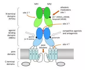
Structural organization of mGluRs Extracellular Ligand binding Domain (EC) • mGluRs are disulfide linked dimers • Two ligand binding sites • Orthosteric: Glutamate, competitive agonists and antagonists (EC domain) • Allosteric : Non-Competitive ligands (TM domain) Glutamate Binding site (Open / Closed) Cysteine-rich Domain (CR) Allosteric Binding site (Active / inactive) Transmembrane Domain (TM)
Available EC domain structures There is no structural data for mGluR6
Functional assay for mGluR6: cAMP assay mGluR6* Adenylyl Cyclase (AC) β γ Forskolin (FK) stimulates cAMP production αo/i* cAMP • mGluR6 is a Gαocoupled receptor • Active (Gαi/o*) negatively regulates AC • inhibits AC activity • decreases cAMP PDE inhibitor (IBMX) blocks cAMP hydrolysis AMP mGluR6 activation decreases cAMP
Effect of agonists on mGluR6 L-glutamate L-ap4 EC50 = 1.2 - 7.5 μM EC50 = 0.1 – 0.3 μM • Determined EC50 matches with the values in literature Stably expressed mGluR6 in HEK293 cells is active
Effect of antagonist on mGluR6 LY341495 • Addition of LY341495 right shifts the dose response curves for • L-AP4 and L-Glu mGluR6 activity is diminished in presence of antagonist
Effect of UBP1112 on mGluR6 • Uninduced • Induced • UBP1112 is reported to be selective antagonist for group III mGluRs UBP1112 appears to be an inverse agonist
Summary of ligand effects on mGluR6 activity: cAMP assay Calcualted EC50 values for agonists is similar to reported values
Modeling and docking studies of orthostericligand binding pocket
Approach Specific Aim 1
Generation of Homology models • Modeller software package • Multiple templates are used to generate average models • Closed mGluR6 • 1EWV_B, 1EWK_B, 1EWT_A, 1EWT_B, 1ISS_A, 1ISS_B • Open mGluR6 and mGluR3 • 1EWV_A, 1EWK_A, 1ISR_A, 2E4U_A, 2E4U_B, 2E4V_A, 2E4V_B, 2E4W_A, 2E4W_B, 2E4X_A, 2E4X_B, 2E4Y_A, 2E4Y_B • Model evaluation • Modeling scores and PROCHECK • Ramachandran plots Models generated: open & closed mGluR6, open mGluR3
Receptor structures for docking Crystal structures used as docking controls
Testing docking approach and receptor models • Primary validation • Verify if ligands from crystal structures when docked recapitulate their binding • Secondary validation • Prepared a database of all the ligands from literature • Docked ligands from database and compare results to experimental binding data
Docking studies • AutoDockVina is used • Rigid docking of receptor, but flexible ligand • Grid box: 30 x 30 x 30 • Exhaustiveness = 64 • Total of 10 docking poses generated • Best hit is the top ranked structures • Successful docking • Top ranked structure docks in binding pocket
Positive controls for docking mGluR1 mGluR3 LYS-409 LYS-389 ARG-68 SER-186 ALA-172 TYR-74 ALA-187 SER-173 Crystal Structure THR-188 THR-174 SER-151 SER-165 LYS-409 LYS-389 ARG-68 SER-186 Docked Structure ALA-172 TYR-74 ALA-187 SER-173 ARG-64 ARG-323 THR-188 THR-174 SER-165 SER-151 -6.2 kcal/mol -6.2 kcal/mol Docking results with glutamate are close to crystal structures
Optimization of Glutamate binding pocket in mGluR6 MODEL OPTIMIZED MODEL ARG-68 SER-155 GLN-64 LYS-400 ASN-281 LYS-312 L-Glu docking -5.7 kcal/mol (Docking pose 3) L-Glu docking -6.1 kcal/mol (Docking pose 2) Optimized model is used for mGluR6 docking studies
Glutamate binding pocket in mGluR6 mGluR6 LYS-400 ARG-68 LYS-409 LYS-389 ALA-175 ARG-68 SER-186 ALA-172 SER-176 LYS-312 TYR-74 ALA-187 Docked Structure SER-173 THR-177 THR-188 THR-174 SER-154 -6.1 kcal/mol SER-151 SER-165 ASN-281 mGluR1 mGluR3 Crystal Structure Glutamate docking pocket in mGluR6 is analogous to that of mGluR1 and3
Docking of dipeptides to closed mGluR6 model • Gly-Glu and Gly-Gln • did not dock • cyclo-Gly-Gln • docked (-6.7 kcal/mol) Gly-L-Glu Gly-L-Gln cyclo-Gly-Gln Cyclo Gly-Gln possible agonist?
Effects of Dipeptides on mGluR6 function Gly-Gln Gly-Glu Cyclo Gly-Gln • cyclo Gly-Gln • No effect • Gly-Glu and Gly-Gln • Antagonist effect Gly-Glu and Gly-Gln have effects comparable to UBP1112 which is an inverse agonist
Positive controls for docking (L-Glu) ALA-187 ALA-187 SER-186 TYR-74 SER-186 TYR-74 THR-188 THR-188 SER-165 SER-165 ASP-208 ASP-208 TYR-74 ASP-218 TYR-74 ASP-218 L-Glu docking has poor binding energy (-4.3 kcal/mol )
Positive controls for docking (S-MCPG) ALA-187 ALA-187 SER-186 TYR-74 SER-186 TYR-74 LYS-409 LYS-409 THR-188 THR-188 SER-165 SER-165 ASP-208 ASP-208 TYR-74 ASP-218 TYR-74 ASP-218 S-MCPG docking does not recapitulate the docking pose seen in the crystal structure
mGluR1: Open (LY341495 docking) LYS-409 TYR-74 SER-186 ALA-187 ALA-187 SER-186 TYR-74 LYS-409 THR-188 THR-188 SER-165 SER-165 ASP-208 ASP-208 TYR-74 ASP-218 TYR-74 ASP-218
Plan for next 3 months • Develop homology model for mGluR6 based on one template alone • Include glutamate in docking • Generate models for all mGluRs which lack crystal strucures • Optimize docking to open structures • Arguslab, Autodock, MOE • Dock ligands from literature and perform comparative analysis • How well do the computational studies correlate with experimental? • What are the key differences between binding pockets of different mGluRs? • Strategy for developing selective ligands
Orhtostericligand binding pocket Group I Group II Group III Green: Identical across all groups; Yellow: Partially identical across groups Red: Different residues within group; None: Different across groups; Grey: Bulky residue in group I and group II not present in group III