Download
1 / 47
770 likes | 2.02k Vues
MEDIASTINAL DISEASES. University Hospital in Łódź Clinic of Thoracic Surgery, General Surgery and Oncology. Author: dr n.med. Sławomir Jabłoński. Mediastinum- anatomical borders: from the top: superior aperture of the thorax, from the bottom: superior surface of the diaphragm,

E N D
MEDIASTINAL DISEASES University Hospital in Łódź Clinic of Thoracic Surgery, General Surgery and Oncology Author: dr n.med. Sławomir Jabłoński
Mediastinum- anatomical borders: • from the top: superior aperture of the thorax, • from the bottom: superior surface of the diaphragm, • from the front: posterior surface of the sternum, • from the back : front surface of the spine and ribs, • from the side : plaques of the mediastinal pleura. Topographically mediastinum is divided into three parts, which include given organs: • upper-anterior: thymus, large vessels, • central: tracheal bifurcation, main bronchi, heart, • posterior: oesophagus, aorta, vagus nerve, sympathetic trunks, lymphatic vessels, azygos vein system.
MEDIASTINAL TUMORS As for histogenesis mediastinal tumors come from different tissues. „ In no other part of human body there are not as many different tumors on such a small space ”. Benszny’ak I. I wsp. Wyd. Akademiai Kiado, Budapest, 1984 Primary mediastinal tumors come from connective tissue, blood and lymphatic vessels. Approximately 55% of tumors are located in an upper-anterior part, 25% in posterior mediastinum and 20% in central mediastinum. Benign tumors of mediastinum are about 60-75% of all tumors. A part of these tumors is located in a characteristic way in this anatomicalarea.
Histopathologic classification of mediastinal tumors - Davis - 1987r. • 1. Neurogenic tumors (p.m.), • 2. Thymic tumors (a.m.), • 3. Lymphomas (c.m), • 4. Embrional tumors (a.m), • 5. Mesenchymal tumors (a.m. c.m.), • 6. Endocrine tumors (a.m.), f.m – anterior mediastinum c.m- central mediastinum p.m- posterior mediastinum
Histopathologic classification of mediastinal tumors - Davis - 1987r. • 7. Primary cancers (c.m.), • 8. Other unclassified tumors, • 9. Cysts: a/ pericardial (c.m.), b/ bronchial (c.m.), c/ from alimentary tube (p.m.), d/ others. Davis R.D.: Ann. Thorac. Surg. 1987, 44, 229.
Mediastinal pseudotumors • Aortic aneurysm, • Heart tumors, • Oesophageal tumors, • Retrosternal goitre, • Inborn Morgagni – Larrey diaphragmatic hernias, • Neoplastic metastases to mediastinal lymphatic nodes.
Frontal mediastinum Frontal mediastinum tumors: • retrosternal and mediastinal goitre • thymomas • embrional tumors (dermatoid cyst, teratomas) • mesenchymal tumors • lymfadenopathies (Hodgkin disease, non-Hodgkin lymphomas) • aneurysms
Mediastinal tumors – clinical picture 80 % of benign tumors – asymptomatic course until the tumor is large and presses other mediastinal structures, 80% of malign tumors – clinical symptoms reveal early in connection to aggressive course of the disease The lack of clinical symptoms is observed most frequently in cysts and benign tumors of posterior mediastinum. Anterior mediastinum tumors due to their location close to different organs cause pressure symptoms quite early.
Mediastinal tumors – clinical picture Most mediastinal tumors cause similar clinical symptoms: TOPICAL SYMPTOMS: • cough, • stridor, • displacement of trachea, • superior caval vein syndrome, • thoracic pain, • pleural exudate, • dysphagia • Horner syndrome, • hoarsness GENERAL SYMPTOMS: • fever • muscle strain • cutaneous rash • pruritus • cancerous cachexia
Mediastinal tumors - diagnosis Diagnostic procedures must provide answers to three basic questions: • Isn’t the observed shadow a vascular structure? • Will a complete surgical treatment be possible ? • Is the surgical treatment necessary?
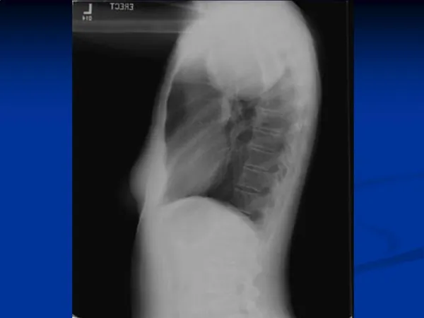
Mediastinal tumors - diagnosis Mediastinum is difficult to be examined phisically and poor clinical symptoms cause that almost 2/3 of all tumors may be difficult to differenciate: Methods of describing the location of tumor in the mediastinum: • Chest X-ray, • Computer tomography with contrast, • NMR examination, • Transesophageal echography (TEE), • Conventional and transesophageal ultrasound examination, • Angiography ( exclusion of aneurysms and inborn malformations of vascular system),
Mediastinal tumors - diagnosis Methods of describing histopathological type of tumor: • Transcutaneous biopsy (controlled by CT or ultrasound – difficult to reach the tumor, no possibility of recognition of proliferation from lymphatic system), • Open biopsy, • Parasternal mediastinotomy, • Carlens’ mediastinoscopy, • Transbronchial biopsy, • Biopsy from above scalene muscle ( Daniels) • Small thoracotomy, • Videothoracoscopy, • Sternotomy with direct histopathological examination,
Mediastinal tumors - diagnosis Examination of neoplastic markers: • S-100 (neurogenic tumors), • CLA, L26, MT1, UCHL1 (lymphomas), • Leu M1 (granulomatosis), • Alphafetoprotein (AFP) Choriongonadotropin (HCG) (embrional tumors).
Mediastinal tumors - treatment THE AIMS OF SURGICAL TREATMENT: • Removal of the tumor (complete operation), • Partial removal of the tumor (cytoreductive procedure – lessening of pressure symptoms) • Histopathological diagnosis, assessment of possible methods of combined treatment
Mediastinal surgery - history • Sauerbruch – 1912 r excision of thymus from cervical approach • Blalock - 26.05.1936r - first excision of thymic tumor from approach by sternotomy • Manteuffel, Bross, Rzepecki, Dębicki - creators of modern Polish thoracic surgery. First operations of thymus excisions from approach by sternotomy burdened high mortality rate - 30%, that is why cervical approach was used for many years.
Mediastinal tumors – surgical treatment OPERATIVE APPROACHES TO MEDIASTINUM: • Cervical approach ( benign thymomas, retrosternal goitre) • Longitudinal sternotomy and partial upper sternotomy (anterior mediastinum tumors, part of anterior mediastinum tumors) • Thoracotomy (posterior and central mediastinum tumors) • Little invasive approach without division of sternum ( cervical-substernal-videothoracoscopic) DIAGNOSTIC APPROACHES: • mediastinoscopy • parasternal mediastinotomy • videothoracoscopy • cervical approach
Mediastinal tumors – surgical tactics • X-ray, CT, NMR, ultrasound examinations possibility of total removal of anterior or posterior mediastinum tumors: Operative treatment (even without histopathological examination) • X-ray, CT, NMR, ultrasound examinations non-operative mediastinal tumor no matter where located or central mediastinum tumors matching enlarged lymphatic nodes: Detailed histopathological examination giving a chance for oncological treatment (chemotherapy, radiotherapy). • Operation reducing the mass of the tumor (cytoreduction) – in some tumors enables application of morw effective combined treatment in less toxic doses.
Non-operative methods of treatment of mediastinum tumors In the past few years there has been an important progress in treatment of many neoplasms including mediastinal tumors with non-surgical methods (chemotherapy, radiotherapy). Assessment of the histopathological type of tumor enables choosing the best method of treatment • Hodgkin’s disease ( from 15% to 75%), • Embryonal tumors ( from 15% to 80%), • Embryonal neurinoma (about 50%). odsetek 5-letnich przeżyć
Operative treatment of mediastinal tumors in Poland • 200 operations per year. • Perioperative mortality = 0-3%. • 78% - radical operations, [0,8%]. • 10% - non-radical operations, [5,7%]. • 12% - exploratory operations, [3,4%].
Neurogenic mediastinal tumors They are the most frequent mediastinal neoplasms (about 20-30%) – benign tumors prevail. Main histopathological types: Tumors originating from neurilemmas: • schwanoma • neurofibroma Tumors originating from ganglion: • neuroblastoma • paraganglioma
Neurogenic mediastinal tumors • Located in posterior mediastinum (80-95%), • 6-10% are malignant tumors requiring combined treatment, • Benign hourglass tumors: posterior access micro-neuro-surgical + anterior access videothoracoscopic.
Thymomas Thymomas– tumors originating from epithelial tissue of thymus ( do not include tumors such as theratomas, lymphomas and carcinoid). Location: anterior mediastinum We identify: • benign hypertrophy of thymus • thymic cysts • thymic carcinoma In some cases they co-exist with (myasthenia gravis) Non-invasive thymus tumors have a distinct capsule. An important criteria of diagnosing aggressive tumors is infiltration of the capsule. This is the basis of IV-stage classification of topical progression of thymomas according to Masaoka). Radical excision of tumor is possible in stage I and II and rarely is III.
MYASTHENIA GRAVIS An autoimmunological disease. Pathomechanism of myasthenia involves creating antibodies against acethylocholin receptor (AchR) located on the membrane of neuromuscular plaque, which leads to disorders in conduction of nerve impulses to the muscles and as a result weakening of muscle strength. The main threat for the patient is respiratory insufficiency due to respiratory muscles disorders. The main part in creating the antibodies against AchR plays the thymus. Myasthenia in thymic tumors: • thymomas are responsible for about 9%-16% of all cases of myasthenia • myasthenia accompanies about 30%-61% of all diagnosed thymomas
Clinical classification of myasthenia according to OSSERMAN and GENKINS
MYASTHENIA GRAVIS DIAGNOSIS: ASSESSMENT OF NEUROMUSCULAR CONDUCTION: • clinical symptoms of muscle strain • test with intravenous injection of Tensilon • examination of neuromuscular conduction (electrophysiological tests) • marking antibodies against AchR receptors METHODS OF THYMUS ILLUSTRATION: • classic and stratified chest X-rays • computer tomography • magnetic resonance
MYASTHENIA GRAVIS PRESERVATIVE TREATMENT: • anticholinesterase medicines (Mestinon, Polstigminum, Metylase) • glucocorticosteroids • immunosuppressive medicines (Cyklofosfamid, Azatiopryna, Cyklosporyna) • immunoglobulins • plasmapheresis OPERATIVE TREATMENT:The procedure of excision of the thymus is called thymectomy. The presence of thymoma or other thymic tumor is an indication to operative treatment. Patients in stage II A and II B with high AchR antibody ratio are qualified to operation.Excision of thymus lessens the symptoms or leads to their complete withdrawal.
METHODS OF THYMECTOMY The aim of thymectomy is to remove the thymus and the adipose tissue of Mediastinum and cervix with ectopic thymical focus ( Masaoki and Jaretzki research) Thymectomy from cervical access ( procedure of a limited completion) Thymectomy from longitudinal sternotomy ( good insight into the madiastinum, disadvantage – extensive wound and operative shock) Maximal thymectomy from 2 accesses: cervical and through sternotomy Expanded cervical thymectomy according to Cooper ( application of suspended surgical hook put on jugular incisure of sternum – better insight into mediastinum) Maximal thymectomy from cervical-substernal-videothoraco- scopic access (allows to remove thymus in a little invasive way without cutting the sternum)
Thymomas – surgical treatment • the best results in case of maximal expanded thymectomy (complete operation) + radiotherapy of mediastinum • indicated even partial excision of thymomas infiltrating neighboring organs (cytoredukctive procedure) + radiotherapy + chemotherapy. • prognosis depends on the type of proliferation (Io-IVo). • IIo i IIIo: 5-year survival in 23-54%. COMPLICATIONS: The most serious complication is respiratory insufficiency due to myasthenic crisis ( up to 20%), sporadically bleeding, infection and instability of sternum, paralysis of phrenic and recurrent laryngeal nerve.
Operative access by longitudinal incision of the sternum ( sternotomia longitudinalis) applied in operative treatmentof anterior mediastinum tumors
Medial longitudinal sternotomy as an operative access in anterior mediastinum tumors
Germ-cell tumors • the most frequently in young males, • benign tumors: only surgical treatment, • malignant tumors: treatment depends on diagnosis ( surgical treatment + combined treatment): - carcinoma embryonale, - teratocarcinoma, - chorioncarcinoma.In case of seminoma good results after radiotherapy • neoplasmatic markers in treatment results monitoring - HCG ( choriongonadothropin), - AFP (alphafetoprotein)
Mesenchymal tumors Tumors originating from connective tissue are: • lipomas, • fibromas, • miomas, • hemangiomas and lymphangiomas • choristomas • chondromas • osteomas Malignant forms of these tumors are rare. Complete surgical excision of the tumor is a method of treatment.
Germ-cell tumors These tumors include all germ layers’ tissues so their cells are able to develop into different tissues and organs. They are about 15% of mediastinal tumors. The tumors are the most frequently solid or cystic and involve different tissues inside (including: hair, teeth, cartilaginous and osseous tissue and others). These are: • teratoma benignum • teratoma malignum • cystis dermoidalis • teratocarcinoma Complete surgical incision of the tumor is a method of treatment. In case of malignant tumors we apply radio and chemotherapy.
Neoplasms from lympharic tissue Lymphatic diseases are sometimes manifested as singular tumors in anterior or medial mediastinum. These include: • Hodgkin’s disease • Non-Hodgkin lymphomas • Lymphatic sarcomas • Castelman’s disease (hypertrophy of mediastinal lymphatic nodes) Lymphomas are a contraindication to operative treatment. If the diagnosis is correct the method of treatment is chemotherapy.
Mediastinal cysts Mediastinal cysts are the effect of developmental disorders in the prenatal period. They are usually asymptomatic. The forms of mediastinal cysts: • pericardial • bronchial • from alimentary tube • pseudocysts Treatment involves surgical removal from sternotomy or thoracotomy access, sometimes they can be removed by means of less invasive methods (videothoracoscopy)
Definitions of ascending goitre in mediastinum Retrosternal goitre: the type of goitre which reaches under the sternal manubrium with its lower ends. It is an extrasternal extension of thyroid gland beginning on the neck, where its blood vessels come from. It descends usually in front of the trachea and the left brachiocephalic vein or laterally to the trachea, rarely behind it and the esophagus. Approximately 10% cases may be located in the posterior part of upper mediastinum, mainly on the right. The frequency of occurrence is: 5%-29%. Mediastinal goitre: it is a kind of goitre which descends its lower ends to the aortic arch or lower, no matter where it starts. It has additional well developed vascular supply in mediastinum. The frequency of occurrence: 0,5% - 3,5% Ectopic goitre: ectopic thyroid tissue without connection to the right thyroid gland Located in mediastinum or other parts of the chast having its own vessel supply.
Mediastinal goitre In mediastinum we can find retrosternal, mediastinal and ectopic goitre. • retrosternal goitre is usually removed from cervical access and only 1-2% may require partial upper sternotomy, • in case of mediastinal goitre where lower ends descend under the sternum sternotomy is necessary, • „Struma aberrans” – very rare, it is necessary to cut the sternum. Thyroid goitreis about 7% of mediastinal tumors. Malignant changes may occur in about 5-15% cases of goitre located in mediastinum.
MEDIASTINAL EMPHYSEMA Pneumomediastinum – mediastinal emphysema (the presence of the air in the mediastinum) Causes : • oesephagus damage • tracheal or bronchial damage • complications of diseases leading to the damage of pulmonary tissue or the bronchial wall • iatrogenic complication after diagnostic or therapeutical puncture of mediastinal structures
MEDIASTINAL EMPHYSEMA CLINICAL SYMPTOMS: • pain located behind the sternum • dyspnoea • anxiety • cervical oedema • neck cracking while palpating • subcutaneous emphysema • tachypnoë • cyanosis • there may be symptoms of pneumothorax or heart tamponade co-existing
MEDIASTINAL EMPHYSEMA TREATMENT : Tentative procedures: • decompressing cervical mediastinotomy (incision of mediastinum over jugular incision of sternum and drainage) • drainage of the pleural cavity with opening of the mediastinum Therapeutical procedures: • debridement of the damage: trachea, bronchi, esophagus • sometimes only mediastinal drainage in case of early diagnosed small esophageal, tracheal or bronchial damages
MEDIASTINITIS Mediastinitis an inflammatory state in loose tissues and mediastinal spaces due to penetrating infection. Severe clinical course is loaded with a high mortality rate. Etiology of mediastinitis: • injuries ( the most frequent cause) • complications of surgical procedures • inflammation descending along fascial spaces in purulent states in oral cavity and neck (periodontal abscesses, purulent tonsillitis, cervical abscesses) • inflammation by circulatory or lymphatic route (rarely)
MEDIASTINITIS DIVISIONS : As for the part of mediastinum affected: • Diffuse mediastinitis • Limited mediastinitisAs for clinical course: • Acute mediastinitis (mediastinitis acuta phlegmonosa) • Mediastinal abscess (abscessus mediastini) • Chronic unspecific mediastinitis (mediastinitis chronica nonspecyfica) • Chronic specific mediastinitis (mediastinistis chronica specyfica)- in tuberculosis and actinomycosis
MEDIASTINITIS CLINICAL SYMPTOMS: • pain behind the sternum • dyspnoe • mobile anxiety • leucocytosis • high temperature • cervical edema • neck cracking while palpating • subcutaneous emphysema • tachypnoë • cyanosis • symptoms of septic shock
MEDIASTINITIS DIAGNOSIS : • clinical picture of mediastinitis • presence of purulent inflammation in cervix, oral cavity • widening of the mediastinal shade on the chest X-rays • leak of the contrast outside the esophagus in case of its perforation • identification of the place of esophagus perforation in endoscopy • visualization of the place of tracheal or bronchial damage in bronchofiberoscopy • presence of fistula in the bronchial stump after pneumonectomy (bronchofiberoscopy)
MEDIASTINITIS TREATMENT : • wide spectrum or guided antibiotic therapy • elimination of the cause of mediastinitis (oesophageal perforatiorrhaphy or operative exclution of oesophagus, maintenance of tracheal or bronchial damage, incision and drainage of oral and cervical abscesses) • effective mediastinal drainage from many access points (cervical, infrasternal, by thoracotomy, exstrapleurally from paravertebral access) • best results when irrigation drainage applied (up-bottom type) –antiseptic solution is given constantly through drain put in the upper part of mediastinum and received through drains in the bottom part of the pleural cavity and under xiphoid process.