Download

1 / 67
670 likes | 825 Vues
BACK PAIN. DR.AFSAR SAYEEDA MRCP(UK) CONSULTANT & HEAD, CTU DEPT OF MEDICINE, KKUH,RIYADH. Epidemiology. 84% of adults experience back pain at some point in their life. - incidence age 35- 55 y.o . - 90% resolve in 6 weeks - 7% become chronic

E N D
BACK PAIN DR.AFSAR SAYEEDA MRCP(UK) CONSULTANT & HEAD, CTU DEPT OF MEDICINE, KKUH,RIYADH
Epidemiology • 84% of adults experience back pain at some point in their life. - incidence age 35- 55 y.o. - 90% resolve in 6 weeks - 7% become chronic - M/ F equally affected • 85% never given precise pathoanatomical dx • 5th Leading reason for medical office visits • 2nd to respiratory illness as reason for symptom-related MD visits
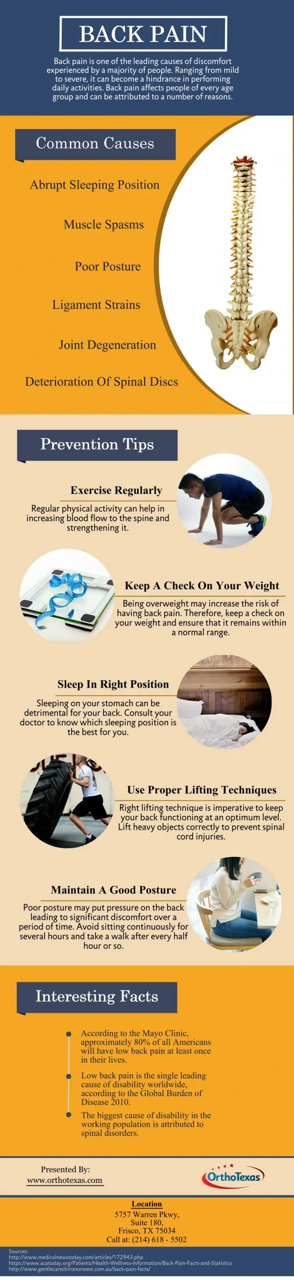
Back Pain in the Primary Care Clinic • 90% of low back pain is “mechanical’ • Injury to muscles, ligaments, bones, disks • For many individuals Spontaneous resolution is the rule. • however, back pain is recurrent or chronic, causing significant pain that interferes with employment and quality of life.
Rarely, acute back pain is a harbinger of serious medical illness, • So don’t miss them! • CAUSES OF NONMECHANICAL/ INFLAMMATORY- • Spondyloarthropathy • Spinal infection • Osteoporosis • Cancer • Other systemic disease • Referred visceral pain
Epidemiology (cont.) • #1 Cause and #1 Cost of work related disability • Healthcare expenditures $100 Billion per year. - $26.3 Billion attributable to back pain
PROGNOSIS GOOD • Acute: 50% are better in 1 week; 90% have resolved within 8 weeks • Chronic: <5% of acute low back pain progresses to chronic pain • Back pain has a substantial impact on lifestyle and quality of life • Psychosocial variables are stronger predictors of long-term disability than anatomic findings found on imaging studies. • predictors of disabling chronic low back pain at one year follow-up--- • maladaptive pain coping behaviors, • functional impairment, • poor general health status, • presence of psychiatric comorbidities, • nonorganic signs
RISK FACTORS FOR ONSET OF BACK PAIN • smoking, • obesity, • older age, • female gender, • physically strenuous work(jobs involving lifting, pulling, or pushing objects of at least 25 pounds) , • jobs involving prolonged periods of standing or walking, especially among women • sedentary work, • psychologically strenuous work, • low educational attainment, • Workers' Compensation insurance, • job dissatisfaction and • psychological factors such as somatization disorder, anxiety, and depression
Important Questions 1. Is systemic disease the cause? 2. Is there social or psycological distress that prolongs or amplifies symptoms? 3. Is there neurologic compromise that requires surgical intervention?
To Answer These Important Questions 1. Careful History and Physical Exam 2. Imaging and Labs WHEN indicated
Clues To Systemic Disease • Age Over 50 years or less than 40 • History of Cancer • Fever • Unexplained Weight Loss • Nighttime pain • Injection Drug Use • Chronic Infection Elsewhere • Duration of pain greater than 1 month • and Quality of Pain -Infection and Cancer not relieved supine • unresponsiveness to previous therapies • h/o inflammatory arthritis elsewhere
Physical Examination • Fever – possible infection • Vertebral tenderness - not specific and not reproducible between examiners • Limited spinal mobility – not specific (may help in planning P.T. • If sciatica or pseudoclaudication present – do straight leg raise • Positive test reproduces the symptoms of sciatica – pain that radiates below the knee (not just back or hamstring) • Ipsilateral test sensitive – not specific: crossed leg is insensitive but highly specific • L-5 / S-1 nerve roots involved in 95% lumbar disc herniations
Imaging • Usually unnecessary & not helpful • Plain Radiography limited to patients with: -findings suggestive of systemic disease -trauma -Age>50years • Failure to improve after 4 to 6 weeks • CT and MRI more sensitive for cancer and infections – also reveal herniation and stenosis • Useful if they have sciatica • Reserve for suspected malignancy, infection or persistent neurologic deficit
Why Not Get Imaging Studies for Acute Back Pain? • Imaging can be misleading: Many abnormalities as common in pain-free individuals as in those with back pain • If under age 60 • Low yield: Unexpected x-ray findings in only 1 of 2,500 patients with back pain • May confuse: Bulging disk in 1 of 3 • Herniated disks in 1 of 5 pain-free individuals If over age 60 and pain free • Herniated disk in 1 of 3 • Bulging disk in 80% • All have age-related disk degeneration • Spinal stenosis in 1 of 5 cases
CT SCAN MRI Shows tumors and soft tissues (e.g., herniated discs) much better than CT scan Almost never an emergency Exception: Cauda equina syndrome • Shows bone (e.g., fractures) very well • Good in acute situations (trauma) • Soft tissues (discs, spinal cord) are poorly visualized • CT-myelogram adds contrast in the CSF and shows the spinal cord and nerves contour better
TERMINOLOGY • terms used to describe conditions related to the back, based upon • radiological findings -(spondylosis, spondylolisthesis, spondylolysis) • physical findings (lumbar lordosis, kyphosis, scoliosis), and • clinical or neurologic features (neurogenic claudication, radiculopathy, sciatica, cauda equina syndrome). • clinical entities that have been associated with low back pain symptoms that are either hard to reliably diagnose or are not clearly associated with symptoms, including the piriformis syndrome, "back mouse," annular tears, and sacroiliac joint dysfunction.
Piriformis syndrome a condition in which the piriformis muscle compresses or irritates the sciatic nerve passing deep or through it. The piriformis muscle is a narrow muscle located in the buttocks. Pain on resisted abduction / external rotation of leg
Sciatica • The sciatic nerve is the longest nerve in your body. • It runs from your spinal cord to your buttock and hip area and down the back of each leg. • Sciatica: pain, numbness, tingling in the distribution of the sciatic nerve, radiating down the posterior or lateral aspect of the leg, usually to the foot or ankle. -herniated disk -foramenal or spinal stenosis -ligamentous hypertrophy -other space filling lesions: cysts, tumor, abscess -viral or immune inflammation -can occur w/ peripheral nerve involvement
LUMBOSACRAL RADICULOPATHY • The clinical presentations vary according to the level of nerve root or roots involved. • The most frequent are the L5 and S1 radiculopathies. • Patients present with pain, sensory loss, weakness, and reflex changes consistent with the nerve root involved.
STRAIGHT LEG RAISING TEST The straight leg raise test is positive if pain in the sciatic distribution is reproduced between 30° and 70° passive flexion of the straight leg. Dorsiflexion of the foot exacerbates the pain
Cauda Equina Syndrome: • Caused by massive midline disc herniation,bony stenosis, or mass compressing cord or( cauda equina bottom-most portion of the spinal canal and spinal nerve roots), • Rare (<.04% of LBP patients). • Needs emergent surgical referral. • Symptoms: bilateral lower extremity weakness, numbness in the groin & saddle area of the perineum, or progressive neurological deficit. • Ask about: • Recent urinary retention (most common) or incontinence? • Fecal incontinence?
LUMBAR SPINAL STENOSIS • local, segmental, or generalized narrowing of the central spinal canal by bone or soft tissue elements, usually bony hypertrophic changes in the facet joints and by thickening of the ligamentumflavum. • Subtle presentation. • Bilateral radicular signs should alert to possibility. • radiation to buttocks, thighs, lower legs • -pain increase with extension (standing, walking- worse on flat) • -pain decrease with flexion (sitting, stooping forward shopping trolley sign) • Can be mistaken for Claudication. neurogenic claudication (pseudo claudication) • 1 or both legs • Admit if progressive / or else CT scan
Management of Spinal Stenosis: Controversial and Evolving • Symptoms of pseudoclaudication without neurologic deficits: • Epidural corticosteroids • Progressive exercise program • Surgical decompression • May relieve leg symptoms • May not relieve back pain • With neurologic deficits: Call the surgeon
Disc Herniation – Physiology Tears in the annulus Herniation of nucleus pulposus
Disc Herniation – Physiology Compression of the nerve root in the foramen leads to pain 98% disc herniations: L4-5; L5-S1 Impairment: Motor and Sensory L5-S1 L5: Weakness of ankle and great toe dorsaflexion S1: Decrease ankle reflex L5 & S1: Sensory loss in the feet
Why Not Get an Operation for a Herniated Disk? • Spontaneous recovery is the rule: 90% resolve over 6 weeks • Predominant symptoms usually leg pain and tingling with less severe or no back pain • Long-term outcome of pain relief no different with or without surgery • only about 10% considered for surgery after 6 weeks • symptomatic and functional outcome sometimes better
Disc Degeneration – Physiology With age and repeated efforts, the lower lumbar discs lose their height and water content (“bone on bone”) Abnormal motion between the bones leads to pain
Low Back Pain - natural history Most episodes of LBP are self limited.(90% in 2wks. – some studies less rapid (2/3 at 7 weeks). These episodes become more frequent with age. LBP is usually due to repeated stress on the lumbar spine over many years (“degeneration”), an acute injury may cause the initiation of pain. LBP is often attributed to disc degeneration, which is the primary target for many diagnostic approaches. the importance of imaging findings associated with disc degeneration (osteophytes, disc narrowing, and herniation) remains unclear. Muscular and ligamentous sources of pain may be equally important.
Waddell Signs For Non-organic Pain • Superficial non-anatomic tenderness • Pain from maneuvers that should notellicit pain • Distraction maneuvers that should ellicit pain BUT don’t • Disturbances not consistent with known patterns of pain • Over-reacting during the exam • Not definitive to rule out organic disease
LBP: Case History 1 • An obese 65-year-old man presents complaining of back pain that began 5 days ago while shoveling snow. The pain becomes worse when he stands • On exam: The spine is nontender, and pain increases with forward bending. Straight leg raising test is negative, and he has no neurologic deficits
Management of Acute LBP: Watchful Waiting • Patient education • Spontaneous recovery is the rule • Those who remain active despite acute pain have less future chronic pain • Exercise has Prevention Power: Muscle strengthening and endurance exercises • Rest: 2 to 3 days or less • Analgesics to permit activity: acetaminophen, NSAIDs, codeine • Reassess if pain worsens
First Episode Acute LBP: Red Flags for Emergent Surgical Consultation • Cauda equina syndrome • Bilateral sciatica, saddle anesthesia, bowel/bladder incontinence • Abdominal aortic aneurysm • Pain pattern is variable • Bruits • +/- pulsatile abdominal mass • Significant neurologic deficit • If they can’t walk, they can’t be sent home
When the Patient Does Not Improve... • The patient returns in 6 weeks because the pain has not decreased. His legs feel “heavy,” and he has had some incontinence in the last week • On exam: He now has bilateral weakness of ankle dorsiflexion, absent ankle jerks, and saddle anesthesia • Diagnosis – Cauda equina syndrome
What Are the Red Flags for Serious Low Back Pain? • Fever, weight loss, night sweats • Acute onset in the elderly • Intractable pain—no improvement in 4 to 6 weeks • Nocturnal pain or increasing pain severity • Morning back stiffness with pain onset before age 40 • Neurologic deficits, bilateral or alternating symptoms. • Sphincter disturbance • Immunosuppression • Infection (current/recent) • Claudication or signs of peripheral ischaemia • History of malignancy
What Should I Be Worried About? • Herniated disk • Spinal stenosis • Cauda equina syndrome • Inflammatory spondyloarthropathy • Spinal infection • Vertebral fracture • Cancer
LBP: Case History 2 • A 32-year-old man complains of severe low back pain of gradual onset over the past few years. The pain is much worse in the morning and gradually decreases during the day. He denies fever or weight loss but does feel fatigued • On exam: There is loss of lumbar lordosis but no focal tenderness or muscle spasm. Lumbar excursion on Schober test is 2 cm. No neurologic deficits
How to Diagnose Inflammatory Back Disease • History • Insidious onset, duration >3 months • Symptoms begin before age 40 • Morning stiffness >1 hour • Activity improves symptoms • Systemic features: Skin, eye, GI, and GU symptoms • Peripheral joint involvement • Infections
How to Diagnose Inflammatory Back Disease (cont’d) • Physical examination • Limited axial motion in all planes • Look for signs of infection • Staph, Pseudomonas, Brucella, and TB • Systemic disease (AS, Reiter’s, psoriasis, IBD) • Ocular inflammation • Mucosal ulcerations • Skin lesions
10 15 cm Testing Spinal Mobility: Schober’s Test • Two midline marks 10 cm apart starting at the posterior superior iliac spine (dimples of Venus) • Premeasure with lumbar spine at maximal flexion • Less than 5 cm difference suggests pathology
Management of Inflammatory Back Pain • Stretching and strengthening exercises • Conditioning exercises to improve cardiopulmonary status • Avoid pillows • NSAIDs • Sulfasalazine • Methotrexate • “biologics”
LBP: Case History 3 • A 40-year-old woman complains of continuous and increasing back pain for 3 months that worsens with movement. She has noted nightly fevers and chills. She is in a methadone maintenance program • On exam she is exquisitely tender over L4 and the right sacroiliac joint with paravertebral muscle spasm. No neurologic deficits. Old needle tracks in both arms • Lab: Hbg 11.5 mg%, WBC 9,000, ESR 80 mm/h
Red Flags for Spinal Infections • Historical clues • Fever, rigors • Source of infection: IV drug abuse, trauma, surgery, dialysis, GU, and skin infection • Physical exam clues • Focal tenderness with muscle spasm • Often cannot bear weight • Needle tracks • Lab clues: Mild anemia, elevated ESR, and/or CRP