Download
1 / 1
10 likes | 296 Vues
Canine Distemper Virus in Raccoons from South Georgia and North Florida Krista A. Cox, Department of Biology Faculty Sponsor: Dr. J. Mitchell Lockhart, Department of Biology. ABSTRACT

E N D
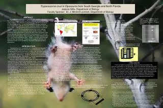
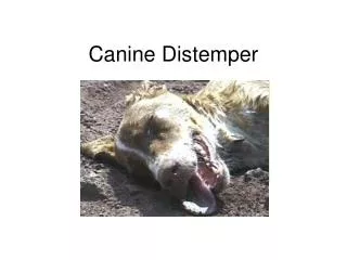
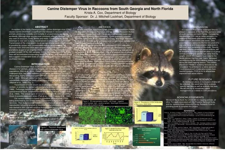
Canine Distemper Virus in Raccoons from South Georgia and North Florida Krista A. Cox, Department of Biology Faculty Sponsor: Dr. J. Mitchell Lockhart, Department of Biology ABSTRACT A member of the family Paramyxoviridae, canine distemper virus (CDV) causes Canine Distemper, a significant viral disease in raccoons and fur animals, producing morbidity and mortality in unvaccinated populations worldwide. Although dogs are the most commonly affected species, CDV is also seen in raccoons, foxes, ferrets and minks. Comparatively, canine distemper is now rare in many industrialized countries due to vaccination. Transmission of the virus occurs via an aerosol-droplet route, direct contact, or possible by contact with contaminated objects. Approximately 365 raccoons from 2003 and 2004 were obtained from three southwest Georgia plantations as part of an ongoing USDA – Wildlife Services bobwhite-quail predator project. Of the 365 samples, 116 (31.8%) tested positive for CDV. Data will be evaluated incorporating season, host sex, and host age. These data suggest considerable levels of canine distemper virus are present in southwest Georgia. METHODS In 2003 and 2004, mesomammalian predators removed from Pebble Hill Plantation, Pinebloom Plantation and Tall Timbers Research Station in southwest Georgia and north Florida were euthanized, frozen and transported to Valdosta State University. These predators included raccoons, opossums, foxes, armadillos, coyotes, feral dogs, feral cats and bobcats. Animals were necropsied and various tissue samples and morphometric data was collected. 365 raccoon serum samples were analyzed for CDV antibodies. Fluorescent antibody slides were commercially obtained from VMRD (Pullman, WA) and consisted of virus-infected mink lung cells grown on the surface of Teflon-masked slides. Fifty µl of raccoon serum diluted 1:8 in phosphate buffered saline was place in designated wells and incubated. Fifty microliters of FITC labeled goat anti-raccoon IgG (Kirkegaard Perry Laboratories) was placed on each well and slides were again incubated. Slides were rinsed, blotted, mounted and were viewed with a microscope at 100X – 250X. Positive samples exhibited a pattern of fluorescence in individual cells with inclusions bodies (Figure 2). DISCUSSION It is apparent that Canine Distemper Virus is present in raccoons of the Red Hills plantation area of south Georgia and north Florida. Considering the effect that CDV has on naïve raccoons, it is surprising that steady prevalence levels were found between plantations and sexes. Data presented here represents the first two years of a four year study and more complex data analysis will occur upon completion of the study. Specific age data, utilizing cementum annuli analysis, will be available in the future and will allow us to group animals according to specific age rather than by weight. USDA-WS trappers have noted to the faculty advisor that they saw “many” deceased raccoons during the 2003 trapping season, but not during the 2004 trapping season. This initially piqued our interest in studying raccoons as we considered the potential for a rabies epizootic. Future analysis will include polymerase chain reaction analysis (PCR) to try to differentiate exposure from active viremia. INTRODUCTION Canine Distemper Virus (CDV) causes distemper, a significant viral disease in fur animals, producing mortality and morbidity in unvaccinated populations (Shin et al., 2004). Closely related to measles virus and rinderpest virus, two other members of the genus Morbillivirus, CDV is a devastating, highly contagious pathogen with a worldwide distribution (Frisk et al., 1999). Although rabies has been more recognized, CDV also has substantially impacted the health of raccoons and wild and domestic canids (Mitchell et al., 1999) (Fig. 1). Transmission of CDV occurs via an aerosol-droplet route, direct contact, or possibly by contact with contaminated objects (Mitchell et al., 1999). Pathologic lesions form and are mostly found in the respiratory and gastrointestinal tracts, lymphoid tissues and central nervous system (Frisk et al., 1999). The clinical signs of raccoons infected with CDV are similar to those of psuedorabies and rabies (Platt et al., 1983; Thawley et al., 1982). Our current research is linked to a predator removal study (PRS) being performed by the United States Department of Agriculture – Wildlife Services. That study involves monitoring the effects of bobwhite quail reproduction following removal of mesomammalian predators. The study was started in 2001 and will continue through 2006 and includes the following cooperative agencies: USDA-WS, The Universityof Georgia, Auburn University, the Jones Ecological Research Center, and Tall TimbersResearch Station.Valdosta State University joined the study in 2003. Theobjective of the present study was to test PRS raccoons for the presence of canine distemper virus specific antibodies via an indirect fluorescent antibody (IFA) assay. RESULTS 47/136 (34.6%) of raccoons tested positive for CDV from Pebble Hill and Pinebloom-East in 2003 (Figure 3). In 2004, 69/229 (30.1%) samples were positive from Pinebloom-West and Tall Timbers. There was no significant difference in CDV prevalence between year of collection (Chi square = 0.58, df=1). A total of 39/116 female raccoons (33.6%) were positive for CDV in 2003-2004 (Figure 4) while 77/229 males (30.9%) were positive for CDV (30.9%). There was no significant difference in CDV prevalence between sex of raccoon (Chi square = 0.16, df=1). No statistical analysis was attempted with month of collection data (Figure 5) or with weight data (Figure 6) as some months and weight groupings had relatively small samples sizes. FUTURE RESEARCH As mentioned above, 2 more years of data will be collected. We would also like to evaluate specific age data as well as antibody titer data. We will also try to incorporate habitat management as it relates to CDV prevalence. ACKNOWLEDGEMENTS We would like to thank the USDA-WS for trapping animals, all other Principals associated with the Predator Removal Study, and the Valdosta State University Faculty Research Fund for providing funds to complete this project. Figure 2. IFA representative results. Left image – negative control sample, right – positive sample. Note bright fluorescence within cells in positive sample. Figure 3. Prevalence of CDV Antibodies In Raccoons, 2003-2004. REFERENCES Frisk, A.L., M. König, A. Moritz, and W. Baumgärtner. 1999. Detection of Canine Distemper Virus nucleoprotein RNA by reverse transcription – PCR using serum, whole blood, and cerebrospinal fluid from dogs with Distemper. Journal of Clinical Microbiology 37: 3634-3643. Mitchell, M.A., L.L. Hungerford, C. Nixon, T. Esker, J. Sullivan, R. Koerkenmeier, and J.P. Pubey. 1999. Serologic survey for selected infectious disease agents in raccoons from Illinois. Journal of Wildlife Diseases 35: 347-355. Parker, R.L., V.J. Cabasso, D.J. Dean, and E.L. Cheatum. 1961. Serologic evidence of certain virus infectious in wild animals. Journal of the American Veterinary Medical Association 138: 437-440. Platt, K.B., D.L. Graham, and R.A. Faaborg. 1983. Pseudorabies: Experimental studies in raccoons with different virus strains. Journal of Wildlife Diseases 19: 297-301. Roscoe, P.E. 1993. Epizootiology of Canine Distemper in New Jersey raccoons. Journal of Wildlife Diseases 29: 390-395. Shin, Y.J., K.O. Cho, H.S. Cho, S.K. Kang, H.J. Kim, Y.H. Kim, H.S. Park, and N.Y. Park. 2004. Comparison of one-step RT-PCR and a nested PCR for the detection of Canine Distemper Virus in clinical samples. Australian Veterinary Journal 82: 6-83. Stanton, J.B., S. Poet, S. Frasca Jr., D. Bienzle, and C.C. Bown. 2002. Development of a semi- nested reverse transcription polymerase chain reaction assay for the retrospective diagnosis of Canine Distemper Virus infection. Journal of Veterinary Diagnostic Investigation 14: 47-52. Thawley, D.G., and J.C. Wright. 1982. Pseudorabies virus infection in raccoons: A Review. Journal of Wildlife Diseases 18: 113-116. Figure 1. Raccoon diagnosed with canine distemper virus