Download
1 / 30
300 likes | 392 Vues
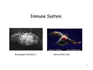
Immune System. Cells and Cell Products That Protect the Body Against Disease. B cells. T cells. Properties of the Immune Response. Specificity: Unique response against each each invader B. Memory: Increased response at next invasion

E N D
Immune System Cells and Cell Products That Protect the Body Against Disease
B cells T cells
Properties of the Immune Response • Specificity: Unique response against each each invader B. Memory: Increased response at next invasion C. Discrimination: Ability to distinguish “self” from “non-self”
Applying Your Knowledge • Specificity • Memory • Discrimination between Self and Non-self Which property of the immune response: • keeps you from developing chicken pox a second time? (Assume you had a severe case at age six.) • helps you successfully recover from a cold? • causes you to reject a donated organ?
Sources and Transport of Immune System Cells • Sites of immune cell production or maturation • bone marrow • spleen • thymus • tonsils • Circulation • lymphatic system • lymph vessels • lymph nodes • bloodstream
Cells of the Immune System T cells carry out cell-cell interactions B cells produce antibodies that bind to antigens on the surface of the invader Macrophages engulf invaders and provide signals for other immune system cells
Activities of Macrophages • Surround invader with plasma membrane and bring it into cell(phagocytosis) • Display antigen on surface by making a complex with MHC protein MHC= major histocompatibility complex, 70 genes that code for cell surface proteins
B Cells Fight Against Viruses and Bacteria • Each B cell carries a unique antibody on its surface • When antigen on surface of invader binds to antibody on B cell, the B cell divides and gives rise to two types of cells --plasma cell: actively secretes antibody into bloodstream --memory B cell: responds on second invasion
Primary Response: initial activation of immune system and destruction of invader Secondary Response: faster and stronger activation of immune system due to memory cells Memory Cells and the Immune Response
Antibody Structure • Antibody has 2 heavy chains and 2 light chains. • Each chain has variable (V) and constant (C) region. • Variable regions bind to antigen. • Constant regions bind to cells or other antibodies.
Gene forconstant regionof light chain Gene forconstant regionof heavy chain Genes forvariable regionof light chain Genes forvariable regionof heavy chain Recombination during Construction of Antibody Genes
Antibodies Mark Invaders for Destruction Phagocytosis by a Macrophage
Applying Your Knowledge • Light Chain • Heavy Chain • Variable Region • Constant Region • Primary Structure • Secondary Structure • Tertiary Structure • Quaternary Structure • Which part of the antibody binds to the antigen? (choices on left) • Which type of protein structure is seen in a functional antibody molecule? (choices on right)
Cytotoxic T cell attacks a cancer cell Cancer cell T cell Classes of T Cells • Helper T cells: activate B cells and cytotoxic T cells • Cytotoxic T cells: rupture infected body cells or cancer cells
1. Macrophage engulfs invader Communication Among Cells 2. Macrophage displays “processed” antigen 7. Dividing B cell gives rise to memory B cells and plasma cells 6. Activated Helper T cell stimulates division of selected B cell 4. Helper T Cell binds to Macrophage 3. Invader binds to B cell that carries antibody matching the antigen 5. Macrophage releases cytokines to activate Helper T cell
Applying Your Knowledge • Macrophage • Plasma Cell • Memory B Cell • Helper T Cell • Cytotoxic T Cell Which immune system cell: • keeps you from developing chicken pox a second time? (Assume you had a severe case at age six.) • helps you successfully recover from a cold? • causes you to reject a donated organ?
Diseases of the Immune System • Inherited Immune Deficiencies eg. SCID: Severe Combined Immune Deficiency B. AIDS: Acquired Immune Deficiency Syndrome C. Autoimmune diseases 1. Scleroderma 2. Rheumatiod arthritis D. Allergies
Allergies Produces antibodies with unique constant region (IgE) IgE antibodies bind to mast cells Antigen binds to B cell and activates it Mast cells release histamines Antigen binds to IgE on mast cells
Envelope(lipid bilayer) CoreProteins HIV ReverseTranscriptase Viral RNA inprotein coat ProteinCoat Glycoproteins
Entry of HIV Retrovirus into Host Cell DNA copy of retroviral genome
Production of New Viral Particles Contains more than one protein product; individual proteins are separated by protease
HIV Binds to CD4 and CCR5 Co-receptors on Helper T cells Individuals that are homozygous for a deletion mutation in the CCR5 gene are resistant to HIV infection.
Use of ELISA to detect Antibodies to HIV ELISA = Enzyme-Linked Immunosorbent Assay Tests serum for the presence of antibodies directed against HIV
Product: Oxidized Salicylate (brown) Substrate: Salicylate (colorless) No brown product formed Anti-IgG with horseradish peroxidase enzyme Anti-IgG with enzyme does not bind No Anti-HIV in serum of uninfected person Anti-HIV in serum of infected person HIV antigen HIV antigen Infected Person Uninfected Person Result Result ELISAProcedure
Instructions for ELISA • Follow ALL directions on pages 90-101 of workbook (summarized in these slides) • Label the vertical side of a microtiter plate as Rows 1, 2, 3, 4 so that there are three wells per row. • Add the antigen: Use a micropipettor and a CLEAN tip to add 0.1 mL (100 uL) of HIV antigen (viral antigens) to all wells. • Incubate at room temperature for 5 minutes then empty the wells with the “flip and bang” method. • Wash the wells: Add PBS buffer with transfer pipette, empty wells with “flip and bang” method.
Instructions for ELISA • Add the test sera: (use clean tip for each) • 100 uL PBS buffer to the three wells in row 1 (negative control). • 100 uL of “+” (positive control: antibody to the HIV antigen) to the three wells in row 2. • 100 uL of Donor Serum 1 to the three wells in row 3. • 100 uL of Donor Serum 2 to the three wells in row 4. • Incubate at 37o (oven on side bench) for 15 minutes then empty the wells with the “flip and bang” method. • Wash the wells: Add PBS buffer with transfer pipette, empty wells with “flip and bang” method.
Instructions for ELISA • Add the 2o Ab: Place 100 uL of the anti-IgG peroxidase conjugate in all 12 wells. • Incubate at 37o (oven on side bench) for 15 minutes then empty the wells with the “flip and bang” method. • Wash the wells: Add PBS buffer with transfer pipette, empty wells with “flip and bang” method. • Add the substrate: Place 100 uL of the substrate in all 12 wells. • Incubate at 37o (oven on side bench) for 5 minutes. • Evaluate the plate for color change