1 / 12

Microscopic images and Cell Specialization

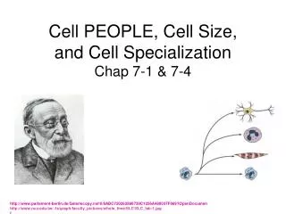
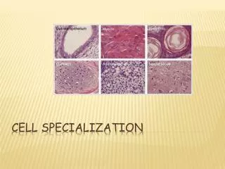
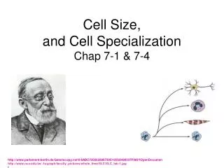
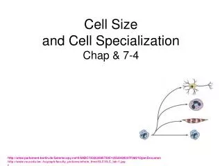
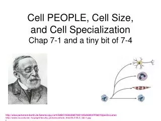
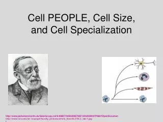
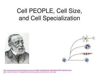
Microscopic images and Cell Specialization. Next time you look under a microscope, observe the relationship between the specimen appearance with unaided eye – and the specimen appearance through the eyepiece. Images appear upside down and backward, compared to slide.

lacy
Télécharger la présentation

Microscopic images and Cell Specialization

An Image/Link below is provided (as is) to download presentation Download Policy: Content on the Website is provided to you AS IS for your information and personal use and may not be sold / licensed / shared on other websites without getting consent from its author. Content is provided to you AS IS for your information and personal use only. Download presentation by click this link. While downloading, if for some reason you are not able to download a presentation, the publisher may have deleted the file from their server. During download, if you can't get a presentation, the file might be deleted by the publisher.

E N D

Presentation Transcript


  1. Microscopic images and Cell Specialization • Next time you look under a microscope, observe the relationship between the specimen appearance with unaided eye – and the specimen appearance through the eyepiece.

  2. Images appear upside down and backward, compared to slide

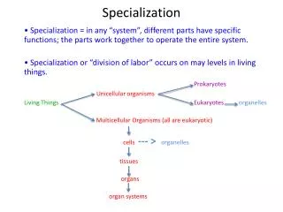
  3. Ask yourself these three questions: • Is it multicellular or unicellular? • What is its function? • How does its shape (its structures) aid in the function?

  4. Amoeba

  5. Animal cells (mitosis)

  6. Muscle Cells

  7. Onion bulb cells

  8. Filamentous Algae

  9. Root cells at edge

  10. Cross section of root

  11. Cross section of plant stem

  12. Cross section of plant leaf

More Related