Download
1 / 37
370 likes | 381 Vues
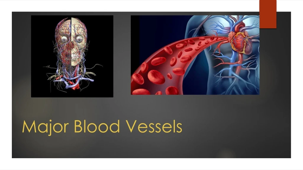
Major Blood Vessels. Vessels. Arteries Veins Capillaries. Structure. Walls contain 3 layers Tunica Externa anchors vessel to surrounding tissues Tunica Media elastic fibers present to allow stretching/recoil Tunica Intima lining of the inner vessel (contains epithelia cells).

E N D
Vessels • Arteries • Veins • Capillaries
Structure • Walls contain 3 layers • Tunica Externa anchors vessel to surrounding tissues • Tunica Media elastic fibers present to allow stretching/recoil • Tunica Intima lining of the inner vessel (contains epithelia cells)
THE ARTERIES THAT SUPPLY THE BRAIN & THE CIRCLE OF WILLIS • Vertebral – spinal cord • Basilar -Spinal cord, Medulla Oblongata, Pons • Posterior cerebral a- Posterior Communicating a • Posterior communicating a • Internal carotid a- anterior half of cerebrum • Middle cerebral a- Mesencephalon, cerebral hemispheres • Anterior cerebral a- frontal & parietal lobes • Anterior communicating a- It communicates both Anterior Cerebral a ALFONSO A. PINO. MD.
The Cerebral Circle • Location- • It encircles the infundibulum of the pituitary gland • Function of The Cerebral Circle: • Because the brain can receive blood from either the • Carotid & theVertebral arteries, it reduces the • possibilities of serious interruption of circulation ALFONSO A. PINO. MD.
THE CEREBRAL ARTERIAL CIRCLE (CIRCLE OF WILLIS) • POSTERIOR CEREBRAL A • POSTERIOR COMMUNICATING • ANTERIOR CEREBRAL A • ANTERIOR COMMUNICATING A ALFONSO A. PINO. MD.
ARTERIES OF THE CHEST AND UPPER LIMB • L & R subclavian a- arms, chest wall, shoulder, back & CNS • Vertebral a- brain & spinal cord • Mammary a (internal thoracic a)- pericardium &anterior wall of the chest • Axillary a- it crosses the axilla to enter the arm • Humeral circumflex a- head of the humerus • Brachial a- upper limb • Radial a-it follows the radius • Ulnar a- it follows the ulna to the wrist • Superficial & deep palmar arches- hand & digital arteries of the thumb & fingers ALFONSO A. PINO. MD.
ABDOMINAL AORTA • Celiac trunk- liver, stomach, spleen • Left gastric a- stomach, esophagus • Splenic a- spleen & arteries to the stomach • Common hepatic a- liver, stomach, gallbladder, duodenum, pancreas • Superior mesenteric a- small intestine, 1st half of large intestine • Renal a- kidneys • Gonadal a- testes (male) / ovaries (female) • Inferior mesenteric a- distal half of the large intestine ALFONSO A. PINO. MD.
ARTERIES OF THE PELVIS & LOWER LIMBS • Common iliac a- • Internal iliac a- urinary bladder, internal & external wall of the pelvis, external genitalia, thigh, uterus & vagina • External iliac a • Femoral a- skin & muscles of the thigh • Popliteal a- back of the knee join • Anterior tibial a- skin & muscles of the anterior leg • Posterior tibial a- posterior surface of the Tibia ALFONSO A. PINO. MD.
VENOUS RETURN FROM THE CRANIUM • Superior sagittal sinus- interior of cerebral hemispheres & choroid plexus • R & L internal jugular v- cranium, face & neck • Temporal v- surface of the head • R & L external jugularv-neck, face, scalp & salivary glands • R & L brachiocephalic v- head , neck & upper extremities. They create the superior v cava ALFONSO A. PINO. MD.
VENOUS RETURN FROM THE UPPER LIMB • Subclavian v-upper limbs • Axillary v- brachial v • Brachial v • Radial v-deep palmar v (radial side of forearm) • Ulnar v-deep palmar v (ulnar side of forearm) • Basilic v-superficial arch (medial surface upper limb) • Cephalic v- superficial arch • Median ante brachial v-superficial arch • Median cubital v- imp-to collect blood samples it is connected to the basilic v • Superficial palmar v • Deep palmar v
VEINS THAT DRAIN THE LOWER LIMB • Common iliac v • R & L internal iliac v- pelvic muscles, skin, urinary & reproductive organs • R&L external iliac v- lower limb • Femoral v • Popliteal v • Posterior tibial v • Anterior tibial v • Peroneal v • Great saphenous v- superficial v of lower limb • Lesser (small) saphenousv- superficial v of leg & foot ALFONSO A. PINO. MD.
Azygous System • Drain the intercostal muscles of the thorax
Pulmonary Circuit • Pulmonary trunk pulmonary arteries lobar arteries (supply the lobes of the lungs where bronchi are • Gasses diffuse across alveoli walls and pulmonary capillaries • Pulmonary capillaries are drained by venules veins pulmonary veins return blood to left atrium
Fetal Circulation • Respiratory and digestive systems are not completely functional • Placenta facilitates exchange of nutrients, waste, and gasses • Umbilical vein carries nutrients and O2 to fetus • Umbilical arteries carry CO2 and waste away to placenta