Download
1 / 87
971 likes | 1.6k Vues
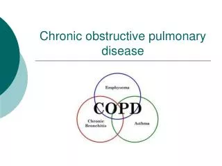
Chronic Obstructive Pulmonary Diseases. Chronic Bronchitis Emphysema Bronchiectasis Bronchial Asthma Small Air way disease Bronchiolitis. Pathogenesis. Prolonged exposure to cigarette smoke and/or other air pollutants. Airway obstruction at times may result in a check-valve mechanism

E N D
Chronic Obstructive Pulmonary Diseases Chronic Bronchitis Emphysema Bronchiectasis Bronchial Asthma Small Air way disease Bronchiolitis
Pathogenesis • Prolonged exposure to cigarette smoke and/or other air pollutants. • Airway obstruction • at times may result in a check-valve mechanism • leading to overdistension and rupture of alveolar septa, especially if the latter are inflamed and exposed to high positive pressure (i.e. barotrauma). • With loss of alveolar surface in emphysema, there is a decrease in surface tension, resulting in expiratory airway collapse. • Additional investigative work continues in an effort to link disease states to pathogenesis
Etiology • By far the most common etiological cause of COPD remains smoking. Even after the client quits smoking, the disease process continues to worsen. • Air pollution • and occupation also play an important role in COPD. • Smog and second-hand smoke contribute to worsening of the disease.
Cont. • Occupational exposure to irritating fumes and dusts may aggravate COPD. • Silicosis and other pneumonoconioses may bring about lung fibrosis and focal emphysema. • Exposure to certain vegetable dusts, such as cotton fiber, molds and fungi in grain dust, may increase airway resistance and sometimes produce permanent respiratory impairment.
Cont. • Exposures to irritating gases, produce pulmonary edema, bronchiolitis and at times permanent parenchymal damage. • Chlorine • Oxides of nitrogen and sulfur, • Repeated bronchopulmonary infections can also intensify the existing pathological changes, playing a role in destruction of lung parenchyma and the progression of COPD.
Chronic Bronchitis • Definition: Chronic bronchitis is a clinical syndrome defined by chronic sputum production • Persistent cough • with sputum production • for at least 3 months • in at least 2 consecutive years • Anatomic site • Bronchus
Morphology • Hyperemia and edema of mm of lungs • Mucinous secretions or casts filling in the air ways • Increase in the size and numbers of mucous secreting glands • Bronchial or bronciolar mucous plugging, fibrosis, inflammation and fibrosis • Squamous metaplaisa or dysplasia of bronchial epithelium
Chronic Bronchitis Pathophysiology Cilia Damaged • Chronic inflammation • Hypertrophy & hyperplasia of bronchial glands that secrete mucus • Increase number of goblet cells • Cilia are destroyed
Chronic Bronchitis Pathophysiology • Narrowing of airway • Starting w/ bronchi smaller airways • airflow resistance • work of breathing • Hypoventilation & CO2 retention hypoxemia & hypercapnea
Chronic Bronchitis Pathophysiology • Bronchospasm often occurs • End result • Hypoxemia • Hypercapnea • Polycythemia (increase RBCs) • Cyanosis • Cor pulmonale (enlargement of right side of heart
Cartilage damage in long standing cases • Degenerative changes, atrophy and loss of bronchial cartilage were common features of most chronic bronchitic specimens • This usually being related to intrinsic changes in the chondrocyte phenotype, including proliferative and matrix-degrading properties. • Mast cells and macrophages were often observed in close association with the bronchial cartilage, • suggesting that inflammatory cells may also contribute to the mechanisms of bronchial cartilage degradation and loss
Inflammatory Cells • Studies show that smokers with symptoms of chronic bronchitis have an increased number of inflammatory cells in theirbronchial glands when compared with asymptomatic smokers. • Thisinflammatory process consists predominantly of neutrophils andmacrophages, and of an increased proportion of CD8+ T-lymphocytes • Mast cells
Mast cells Lymphocytes Neutrophils Macrophages Macrophages, Mast cells, Lymphocytes and Neutrophils
Neutrophils Lymphocytes Mast cells Macrophages
Thisinflammatory process consists predominantly of neutrophils andmacrophages, and of an increased proportion of CD8+ T-lymphocytes and Mast Cells
Pathogenesis • Chronic irritation of airways • Smoking • Dust • Air pollutants etc • The major risk factor for the developmentof chronic bronchitis is cigarette smoking • Infective agents Secondary factor • These irritants cause • Hypersecretions of mucous • Subsequent hypertrophy of mucous glands • Metaplasia • inflammation
Emphysema • Pulmonary emphysema is described in clinical, radiological and physiologic terms, but the condition is best defined morphologically • Definition:Abnormal enlargement of airspaces distal to the terminal bronchioles with destruction of their wall • It is characterized by destruction and enlargement of alveoli
Cont. • Although the normal lung has about 35,000 terminal bronchioles and their total internal cross-sectional area is at least 40 times as great as that of the lobar bronchi but the bronchioles are more delicate and vulnerable.
Cont • Bronchioles may be obstructed partially or completely, temporarily or permanently, by thickening of their walls, by collapse due to loss of elasticity of the surrounding parenchyma, or by influx of exudates.
Cont • In advanced emphysema, the lungs are large, pale, and relatively bloodless. • They do not readily collapse. • They many contain many superficial blebs or bullae, which occasionally are huge. • The right ventricle of the heart is often enlarged (cor pulmonale), reflecting pulmonary arterial hypertension.
Emphysema: Pathophysiology • Structural changes • Hyperinflation of alveoli • Destruction of alveolar & alveolar-capillary walls • Small airways narrow • Lung elasticity decreases
Emphysema • Loss of Lung Surface Area for Gas Exchange and Oxygen Transport • Loss of Lung Surface area is due to death of Lung Endothelial Cells • Polycyclic aromatic hydrocarbons (PAHs) in Cigarette Smoke and in Environmental Pollution may cause endothelial cell death
Emphysema: Pathophysiology • Mechanisms of structural change • Obstruction of small bronchioles • Proteolytic enzymes destroy alveolar tissue • Elastin & collagen are destroyed • Support structure is destroyed • “paper bag” lungs
Emphysema: Pathophysiology • The end result: • Alveoli lose elastic recoil, then distend, & eventually blow out. • Small airways collapse or narrow • Air trapping • Hyperinflation • Decreased surface area for ventilation
Emphysema: Clinical Manifestations • Early stages • Dyspnea • Non productive cough • Diaphragm flattens • A-P diameter increases • “Barrel chest” • Hypoxemia may occur • Increased respiratory rate • Respiratory alkalosis • Prolonged expiratory phase • Tripod position
Emphysema: Clinical Manifestations • Later stages • Hypercapnea • Purse-lip breathing • Muscle retraction • Use of accessory muscles to breathe • Underweight • No appetite & increase breathing workload • Loss of subcutaneous fat • Lung sounds diminished
Clinical Manifestations • Pulmonary function • residual volume, lung capacity, DECREASED FEV1, vital capacity maybe normal • Arterial blood gases • Normal in moderate disease • May develop respiratory alkalosis • Later: hypercapnia and respiratory acidosis • Chest x-ray • Flattened diaphragm • hyperinflation
Classification • Anatomical Distribution • Cetriacinar • Panacinar • Paraseptal • Irregular
Pathogenesis • Protease anti Protease Theory • Hereditary deficiency of the major protease inhibitors • Laurell and Eriksson –1963 – deficiency of α1-antitrypsin and emphysema • Pollutants in environment • smoking of cigarettes • exhalations from cars, dust from grain • Recruitment of neutrophils • stimulation of macrophages and epithelial cells to produce TNF- α, IL-8 and LTB4 • Elastase release from leukocytes and tissue macrophages • Inactivation of α1-antitrypsin by oxidants in tobacco smoke or free radicals released from the neutrophils
Diseases in which misfolding of a given protein results in improper trafficking
Diseases in which misfolding of a given protein results in improper trafficking polymers of alpha1 antitrypsin molecules will form. These will be retained in the liver (where alpha1 antitrypsin is produced) and will not reach the lungs
hyper aeration increased lung lucency decreased vessel size Flattening of the Diaphragm Increased Lung Height Decreased Peripheral Vascular Markings
Chest X-Ray Changes in Emphysema: • The two primary findings are: • Increased Lung Volumes, i.e., hyperaeration or overinflation. • Lung Destruction (bullae or decreased vascularity which gives increased lung lucency and decreased vessel size ). • In end-stage disease, emphysema and chronic obstructive pulmonary disease can result in pulmonary arterial hypertension and eventually cor pulmonale (cp) - which causes an increase in the pulmonary arterial size, as well as right ventricular enlargement and cardiomegaly.
Morphology Centrilobular Emphysema Panlobular Emphysema
Paraseptal Peripheral alveolar damage
Other types of Emphysema 1 Bullous Emphysema (also known as Bullous Lung Disease):Bullous emphysema is so named when there are multiple large bullae associated with a compromise in pulmonary function. It is usually associated with concomitant emphysema, although occasionally, it can be familial
Bullous Emphysema • Subpleural Type: These subpleural bullae contain only gas with no alveolar remnants or blood vessels. They are often located in the apex of the upper lung zone, and along the costophrenic rim of the middle lobe and lingua, but may be seen in the vicinity of parenchymal scars. • Superficial Type: These bullae are found along the anterior edge of the upper and/or middle lung zones, or lingula, and over the diaphragms. They contains blood vessels and strands of partially-destroyed lung. • Deep Type: These bullae are found within the lung substance and contain strands of partially-destroyed lung tissue and blood vessels.
2.Giant Bullous Emphysema: ("Vanishing Lung Syndrome" or "Primary Bullous Disease of the Lungs") • It is usually associated with young males who show large progressive upper lung zone bullae that are often asymmetric. • The giant bullous lesions occupy greater than or equal to one-third of the hemithorax.
Cont. 3.Focal Dust Emphysema ( This is focal emphysema surrounding silicotic nodules. • 4.Congenital Lobar Emphysema (CLE)5.Interstitial Emphysema
Bronchiectasis • Bronchiectasis is a chronic lung disease that is characterized by permanent dilatation of the bronchi and fibrosis of the lung. • It is defined as the pathological, irreversible dilation of bronchi , due to destruction of the bronchilal walls and their supporting tissues • It is highly associated with chronic bacterial infection • Often looked at, as the final common pathway of many injurious processes
Cont. • Bronchiectasis , although uncommon,bears the potential to cause severe illness , including repeated respiratory infections , disabling cough, purulent sputum, shortness of breath, • chest pain and occasionally hemoptysis, with significant impact on the health and the quality of life of the affected person
Types of Bronchiectasis • Bronchiectasis means irreversible dilation and distortion of the bronchi and bronchioles. • Pathologically, bronchiectasis can be divided into four types