Download
1 / 20
210 likes | 421 Vues
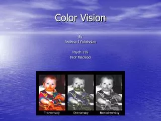
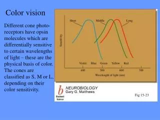
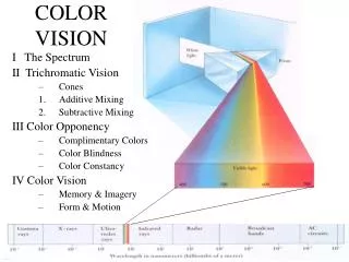
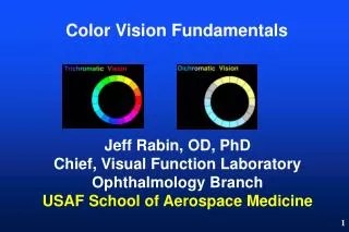
Color vision. Outlines. What is color Retinal circuitry for color vision Several ways to label retinal cells. Color history: discovery of color. Isaac Newton. Color history: discovery of color. Reflection makes colors. Retina anatomy of human being. Outer nuclear layer.

E N D
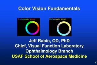
Outlines • What is color • Retinal circuitry for color vision • Several ways to label retinal cells
Color history: discovery of color Isaac Newton
Retina anatomy of human being Outer nuclear layer Outer plexiform layer Inner nuclear layer Inner plexiform layer Ganglion cell layer
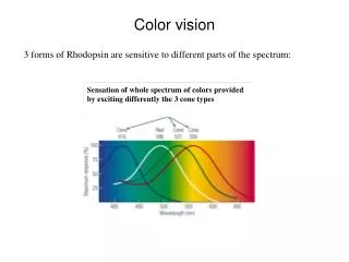
Cone mosaic in human retina Immunohist-ochemistry (IHC) S-cone L-cone S-cone M-cone L-cone or M-cone
Strategy to see color • Principle of Univariance: any single photopigment is “colour blind”. Given proper combination of wavelength and intensity, and can trigger same response in M-cone. • Color-coding ganglion cells receive antagonistic input from cones: red-green; yellow-blue
Color opponency mechanism Red-greenopponent cells: midget ganglion cells Blue-yellowopponent cells: small bistratified ganglion cells 1111 Review by Dacey, 2000
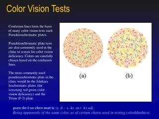
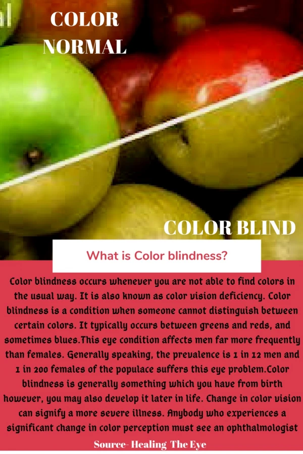
Dichromats Review of Masland, 2012
S/M opponent ganglion cells in retina of guinea pig a dichromatic mammal Dye iontophoresis
Dye iontophoresis What is ? To dye fill a cell: visualize the cell via quick and rough nucleus staining A microelectrode filled with fluorescent dye is placed carefully in contact with the soma Break into the cell and apply current. “injecting fluorescent dye into the cell electronically”. How big is 10μm? The width (diameter) of a human hair ranges from 0.017 to 0.181 mm.
Movie showing iontophoresis of dye into retinal ganglion cell
Summary of cell labeling techniques Immunocytochemistry Dye-filling 3. Genetic tags: GFP, YFP…
History of genetic tag: GFP 2008 Nobel prize winners Osamu Shimomura, Martin Chalfie, and Roger Tsien,
Transgenic lines that mark RGC subtypes Genetic tags: YFP
The End “Most of my knowledge came from self-study,” says Dr. Shimomura. “If you find an interesting subject, study it through to the finish. If you confront difficulties, overcome them. Don’t be discouraged. There are always difficulties in research.”