Download
1 / 33
390 likes | 785 Vues
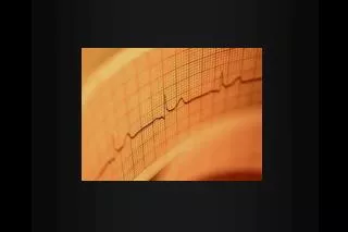
ECG interpretation for beginners Part 4 – Acute coronary syndromes. Paul Williams Cardiology Specialist Registrar. Normal ECG!. MI diagnosis. Use your system Don’t forget rate, rhythm etc. Need to have basic understanding of: Pathology of heart attacks

E N D
ECG interpretation for beginnersPart 4 – Acute coronary syndromes Paul Williams Cardiology Specialist Registrar
MI diagnosis • Use your system • Don’t forget rate, rhythm etc. • Need to have basic understanding of: • Pathology of heart attacks • Coronary arteries and regions of heart
Coronary arteries • 2 coronary arteries come off aorta • Total of 3 main coronary arteries • LCA • LMS branches into: • Left anterior descending (LAD) • Circumflex (Cx) • RCA
Left ventricle supply • LAD – Supplies anterior wall, septum +- lateral walls(60%) • Cx – Supplies lateral wall (15%) • RCA – Supplies inferior and posterior walls (25%). Also supplies RV & conducting tisse
Septal Lateral Anterior Lateral Inferior
Other territories • Inferior MI – can have RV involvement • RV leads - V4R • Posterior MI – Usually ST depression V1-V3
The hallmark of acute ischaemia is ST segment shift • ST elevation = complete blockage = STEMI • ST depression = partial blockage = NSTEMI/USA • Generally only occurs when patient has symptoms: ACS are dynamic • If real, usually have changes in contiguous leads
STEMI • Occluded coronary artery • Emergency = myocardium is dying!
STEMI • Changes evolve: • Often “hyperacute” T waves initially • T wave inversion • Q waves • Dynamic - repeat ECGs if not sure • What territory is it? • Two contiguous leads • Can get reciprocal ST depression • Remember posterior & RV involvement
Differential • Pericarditis • Widespread concave upsloping ST depression • Would involve multiple coronary arteries if MI • PR depression (II) • Look at the patient – common sense
Management of STEMI • ABC • Cardiac monitor (can go into VF) • Analgesia • Aspirin • Clopidogrel • Reperfusion therapy • Thrombolysis • Primary PCI • Medical Rx
Septal Lateral Anterior Lateral Inferior
Old MIs • Old STEMIs can leave permanent Q waves • Territories are the same (anterior, inferior lateral etc.) • Poor R wave progression can also indicate an old anterior STEMI
ST depression • Often get T wave inversion as well • Remember your territories • Generally ST depression only occurs during acute ischaemia • Differential • Digoxin (downsloping lateral: V4-V6, I, aVL) • LVH (downsloping lateral)
Management of NSTEMI/USA • ABC • Cardiac monitor • Analgesia • Initial medical Rx • Aspirin • Clopidogrel • Beta-blocker • Statin • LMWH • IP angiography
Question 1 • What are the ECG abnormalities? • What is the differential?
Question 2 • What are the ECG abnormalities? • What sort of ACS? • What territory is affected?
Question 3 • What are the ECG abnormalities • What sort of ACS? • What territory?
Question 4 • What are the ECG abnormalities? • Give 3 possible differentials
Question 5 • What are the ECG abnormalities? • What sort of ACS? • What territory?