Download
1 / 17
280 likes | 1.37k Vues
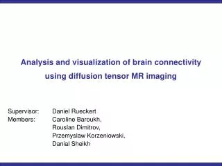
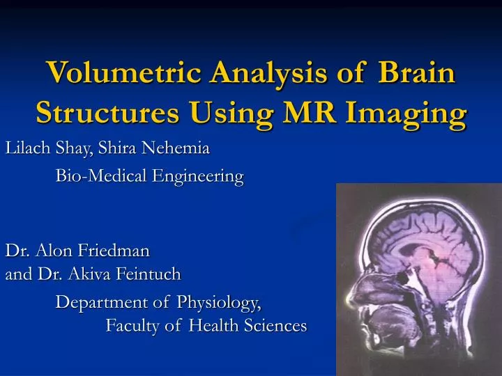
Volumetric Analysis of Brain Structures Using MR Imaging. Lilach Shay, Shira Nehemia Bio-Medical Engineering Dr. Alon Friedman and Dr. Akiva Feintuch Department of Physiology, Faculty of Health Sciences. Basic Principles of MRI. The Human body mainly consist of water

E N D
Volumetric Analysis of Brain Structures Using MR Imaging Lilach Shay, Shira Nehemia Bio-Medical Engineering Dr. Alon Friedman and Dr. Akiva Feintuch Department of Physiology, Faculty of Health Sciences
Basic Principles of MRI • The Human body mainly consist of water • The concept of MRI is based on the fact that hydrogen nuclei have magnetic properties, called nuclear spin • Macroscopic, they behave like tiny rotating magnets, represented by vectors www.e-mri.org
Magnetization Vector • Within a large external magnetic field (called B0), nuclear spins align with the external field. Most of the spins align with the field (parallel) and some align against the field (anti-parallel) www.e-mri.org www.simplyphysics.com
Larmor Frequency • The resonance frequency, called Larmor frequency (ω0) or precessional frequency, is proportional to the main magnetic field strength External magnetic field Larmor Frequency Geomagnetic ratio andis a constant unique to every atom (H+=42.56 MHz/Tesla) www.e-mri.org
Basic principles of MRI A magnet is a dipole and it can be represented by a magnetic vector. A moving magnetic field induces a current in a coil. For example, the rotating magnet below induces a sinusoidal current that can be recorded. www.e-mri.org
Volume and Atrophy • Quantitative volume measurements of specific brain regions are essential for research of brain diseases and may prove of diagnostic value (e.g. neurodegenerative diseases). Insight into pathological mechanisms Diagnosis Monitor treatment efficacy Decision upon treatment
Hippocampus and Cerebral Cortex • Takes part in memory and spatial navigation • Consists of a mixture of grey matter and white matter Schmolesky M., 2000 http://www.thomaskoenig.ch/Lester/ibaspm.htm • Outermost thin layer of grey matter in the brain • Many brain functions: memory, attention, perceptual awareness, language and consciousness
Difficulties of Brain Volume Evaluation • Signal intensity of grey matter and white matter vary within the brain • Contrast of white - grey matter is relatively dim • Partial volume effect • Artifacts
1. Grey level thresholding 2. “Edges” method , - checking neighbor voxels 3. Statistical method - Bayes correction 4. The manual segmentation Review of Segmentation Methods
Review of segmentation methods Bayes law The likelihood component of the tissue The label probability The probability to get a specific grey level value Thacker et al., 2004
Literature review • Review of software packages: 1. MRIcro - -converts the data from slices of 2-D matrixes into one matrix only (3-D) -the MRI data viewer. Up until now
2. SPM- (Statistical Parametric Mapping) .a MATLAB software package. Allows us to perform spatial normalization and smoothing (Bayes correction) of the data. Review of Software Packages
Performing Brain MRI Scans We have performed 2 MRI scans Different sequences: - T1: Recovery of longitudinal magnetization - T2: Decay of transverse magnetization - IR (Inversion Recovery) - FLAIR (Fast fluid attenuated inversion recovery) - Grey Matter only Computer Vision Laboratory at UMass,2003
Data Analyses Writing a MATLAB scripts that: 1. analyzes SPM- generated output 2. produces a mask of Grey Matter 3. Segmentation of Grey Matter applied on T1- weighted MRI
Up until now Segmentation of Grey Matter applied on T1- weighted MRI
Future Tasks Choosing the optimal sequence (minimal error) for grey matter segmentation. Validating the developed tools by measuring grey matter volume in a sample of controls and neurological patients. Develop mathematical tools for delineating the cerebral cortex and the hippocampus from sub- cortical grey matter.
The End Questions ?