Download
1 / 38
380 likes | 572 Vues
Introduction and Classification of Anaemia PALI Haematology Session 3 rd October 2012 Christopher Mullen. Overview of Presentation. Introduction Classification The Hypochromic Anaemias Megaloblastic and other macrocytic anaemias Haemolytic anaemias Genetic disorders of haemoglobin

E N D
Introduction and Classification of Anaemia PALI Haematology Session 3rd October 2012 Christopher Mullen
Overview of Presentation • Introduction • Classification • The Hypochromic Anaemias • Megaloblastic and other macrocytic anaemias • Haemolytic anaemias • Genetic disorders of haemoglobin • Investigation of the anaemic patient • Exam paper cases
Introduction • Reduction in haemoglobin/red cell concentration in the blood relative to the patient’s age and sex.
General Clinical Features • Prevalent worldwide, especially among females and children • A reduction in the blood’s oxygen carrying capacity leading to tissue hypoxia • Clinical features relate to compensatory mechanisms • Cardiovascular: palpitations, chest pain, tachycardia, intermittent claudication • Skin: vasoconstriction and redistribution of blood flow
Classification • Morphological classification • Dependent on red cell indices i.e. MCV and MCH/MCHC • Macrocytic vs. normocytic vs. microcytic • Hypochromia vs. normochromia • Aetiological classification • Haemorrhage • Haemolytic • Insufficient/ineffective haemopoiesis
The Hypochromic Anaemias Differential Diagnosis • Iron deficiency • The most common cause of anaemia worldwide • Hypochromic, microcytic anaemia (↓MCV, ↓MCHC) • Thalassaemia • Sideroblastic anaemia • Lead poisoning • Anaemia of chronic disease (sometimes) • Acronym - TAILS
Iron Deficiency Anaemia Clinical Features • Pallor of skin and mucous membranes • Painless glossitis • Angular stomatitis • Koilonychia
Aetiology Aetiology • Chronic blood loss – uterine and gastrointestinal • Dietary deficiency (rarely alone) • Increased demand – pregnancy, lactation, infancy etc. • Gastrointestinal disease e.g. coeliac, atrophic gastritis, gastrectomy • Helminth infections
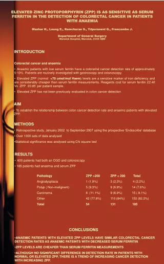
Investigations Iron deficiency anaemia is not a diagnosis – an underlying cause must be sought • Blood film • Iron studies – increased total iron binding capacity (TIBC), low serum ferritin • Men and post-menopausal women GI blood loss occult blood testing, endoscopy if suspicious • Uncommon causes – CXR, stool microscopy, cell autoantibodies
Management • Identify and treat underlying cause(s) • Ferrous sulphate • Parenteral iron where appropriate e.g. coeliac disease, Crohn’s disease
Anaemia of Chronic Disease • Associated with a variety of malignant and inflammatory diseases • Normocytic and normochromic (or slightly microcytic anaemia) • Cytokines reduced red cell lifespan and impair haemopoesis
Macrocytic anaemias • MCV abnormally large (>98 fL) • Alcohol • Liver disease • Hypothyroidism • Myeloma and paraproteinaemia • Myelodysplasia • Reticulocytosis • Pregnancy • Cytotoxic drugs e.g. azothioprine
Megaloblastic anaemia • Immature red cells display delayed nucleus maturation • Due to defective DNA synthesis • Nearly always caused by folate/B12 deficiency
B12 deficiency • Absorbed in terminal ileum (requires intrinsic factor) • Acts as a co-enzyme • Deficiency takes 2 years to develop • Pernicious anaemia – associated with autoimmune diseases. Autoimmune attack against parietal cells/intrinsic factor
Folate Deficiency • Required for synthesis of thymidine monophosphate (dTMP) • Causes • Poor diet • Increased demand – pregnancy (NTDs) • Haematological disease • Inflammatory disease • Malabsorption
Clinical Features • Lemon skin (jaundice and pallor) • Glossitis • Subacute combined degeneration of the cord in severe B12 deficiency • Macrocytic anaemia • Mildly reduced WCC and platelets • Hypersegmented neutrophils • Raised unconjugated bilirubin and LDH
Further Investigations • Serum assays for folate and B12 • Parietal cell and intrinsic factor antibodies • Intestinal biopsy, anti-TG if suspicious for coeliac disease
Haemolytic anaemias • Anaemia due to increased red cell destruction • Can be hereditary or acquired • Hereditary are due to intrinsic defects within red cells • Acquired are due to changes in red cell environment
Classification Hereditary Acquired Immune Autoimmune warm Autoimmune cold Drug-associated Transfusion-associated Infections Malaria Secondary to liver and renal disease PNH • Membrane defects • Hereditary spherocytosis • Hereditary elliptocytosis • Enzyme defects • Glucose-6-phosphate dehydrogenase deficiency • Pyruvate kinase deficiency • Defects in haemoglobin • Haemoglobin C, haemoglobin S, unstable haemoglobin
Clinical Features • Pallor of mucous membranes • Jaundice • Splenomegaly • Damaged red cells on blood film • Erythroid hyperplasia of bone marrow • Bilirubinaemia and increased urinary urobilinogen
Chemical Features of Intravascular Haemolysis Intravascular Haemolysis Extravascular Haemolysis Red cells are broken down by macrophages or the reticuloendothelial system • Breakdown of red blood cells in the circulation • Haemoglobin is released (haemoglobinaemia) • Haptoglobins are saturated • Haemoglobinuria • Haemosiderinuria • Methalbuminaemia
Genetic disorder • Reduced rate of synthesis of α or β globin chains • Common in Mediterranean and South-East Asia • Varies depending on number of genes rendered inactive • Marrow hyperplasia in β-thalassaemia gives rise to characteristic facies • Diagnosed by haemoglobin electrophoresis/high performance liquid chromatography
Genetic disorders of haemoglobin – Sickle Cell Anaemia • Inheritance of the sickle β-globin gene • Homozygotes (Hb SS) are the most common severely affected patients • Severe haemolytic anaemia associated with “crises” • Diagnosed via haemoglobin electrophoresis
You are a GP trainee and Mrs J, a 24 year old lady, presents to the clinic. She has just moved to your area, her notes haven’t arrived at the surgery, and she is complaining of tiredness. She doesn’t wish to be fully examined but clinically you suspect anaemia and you arrange a full blood count, results of which are shown below. She returns to health centre and this time you notice that she is also slightly icteric. Urine analysis shows urobilinogen but no bilirubin. There is no glycosuria, haematuria or pyuria. The serum bilirubin concentration is 65 Kmols/l (normal range 15 – 22 Kmols/litre).
Apart from investigations for haemolysis, list two other investigations, explaining your reason for doing the test to help elucidate the cause for producing the increased MCV (2 marks) • Apart from the results in the previous slide, list two biochemical or haematological abnormalities that may occur in haemolysis
Explain in less than fifty words, why an increase in serum bilirubin will not lead to increased renal excretion of bilirubin. (2 marks) • List two defects in the red cells which can cause haemolysis and give one example of each (2 marks)
It transpires that Mrs J had a splenectomy for this problem as a child and that she subsequently had no follow-up or treatment after this procedure. List two organisms you would wish to vaccinate against. (2 marks) • List two pieces of advice you would wish to instigate in patients following a splenectomy. (2 marks)
A 32 year old woman, who is a mother of four children under aged 6 years, presents with increasing fatigue and shortness of breath over recent months. She has no significant past medical history. You find her to be pale with: Hb 6.9 g/dl (Reference range 11-13 g/dl), MCV 63fl (Reference range 78-96 fl), MCH 24 pg (Reference range 27-32pg)
What name is given to this blood picture? (2 marks) • Microcytichypochromic anaemia • What is the most likely haematological disorder in this lady? (1 mark) • Iron deficiency anaemia
List two possible significant factors underlying in this patient. (2 marks) • What arterial PO2 would you expect? (1 mark)
You examine the blood report to find a reticulocyte count? What are reticulocytes? (1 mark) • What is the significance of a normal result? (1 mark)
On further questioning, you discover this patient’s ethnic background is South Asian. • What co-existing blood condition does this patient have? How would you test for this? (2 marks)
ABurkitt’s Lymphoma • B Chronic Lymphoid Leukaemia • C Chronic Myeloid Leukaemia • D Depression • E Hodgkin’s Lymphoma • F Pernicious Anaemia • GPolycythaemia Vera • H Sickle Cell Anaemia • ISideroblastic Anaemia • J Thalassaemia • A 65-year-old lady presents to her GP with a 3-month history of vertigo, tinnitus and visual disturbance. She admits to feeling “a bit down” and the GP decides to carry out some routine bloods. A week later she returns and you note that her blood results show a raised high haemoglobin and a raised pack cell volume and red blood cell count.
A 12-year-old girl of Nigerian descent and with a known blood disorder presents to A&E with a two-day history of dyspnoea, cough and fever. You order several investigations and note that she has a Hb of 6g/dl (reference range 11.5 – 1.35 g/dl) and a chest X-ray showing pulmonary infiltrates. • ABurkitt’s Lymphoma • B Chronic Lymphoid Leukaemia • C Chronic Myeloid Leukaemia • D Depression • E Hodgkin’s Lymphoma • F Pernicious Anaemia • GPolycythaemia Vera • H Sickle Cell Anaemia • ISideroblastic Anaemia • J Thalassaemia
A 27-year-old man presents with a two-month history of pruritis, fatigue and weight loss. On questioning he admits that whenever he drinks alcohol, he experiences bone pain. On examination he has a rubbery non-tender submandibular lymph node. He has never had infectious mononucleosis. • ABurkitt’s Lymphoma • B Chronic Lymphoid Leukaemia • C Chronic Myeloid Leukaemia • D Depression • E Hodgkin’s Lymphoma • F Pernicious Anaemia • GPolycythaemia Vera • H Sickle Cell Anaemia • ISideroblastic Anaemia • J Thalassaemia
A 68-year-old woman presents with a history of bruising, bone pain and lymphadenopathy. Unbeknownst to the consultant, this patient has a (t9,22) mutation known as the Philadelphia Chromosome. On examination the consultant finds a massively enlarged spleen. • ABurkitt’s Lymphoma • B Chronic Lymphoid Leukaemia • C Chronic Myeloid Leukaemia • D Depression • E Hodgkin’s Lymphoma • F Pernicious Anaemia • GPolycythaemia Vera • H Sickle Cell Anaemia • ISideroblastic Anaemia • J Thalassaemia
A 37 year old lady with known hypothyroidism presents to you with fatigue, dyspnoea and palpitations. You note that she is pale and tachycardic. Routine bloods show a macrocytic anaemia. You suspect that this is caused by her hypothyroidism. You find a positive Schilling’s test. • ABurkitt’s Lymphoma • B Chronic Lymphoid Leukaemia • C Chronic Myeloid Leukaemia • D Depression • E Hodgkin’s Lymphoma • F Pernicious Anaemia • GPolycythaemia Vera • H Sickle Cell Anaemia • ISideroblastic Anaemia • J Thalassaemia