Download
1 / 75
750 likes | 1.19k Vues
Chapter 42: Gas Exchange and Circulatory System. Presented by: McQuade and Verpooten. Circulatory Systems Reflect Phylogeny. In unicellular organisms Exchanges occur directly with the environment For most of the cells making up multicellular organisms

E N D
Chapter 42: Gas Exchange and Circulatory System Presented by: McQuade and Verpooten
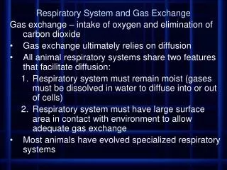
Circulatory Systems Reflect Phylogeny • In unicellular organisms • Exchanges occur directly with the environment • For most of the cells making up multicellular organisms • Direct exchange with the environment is not possible
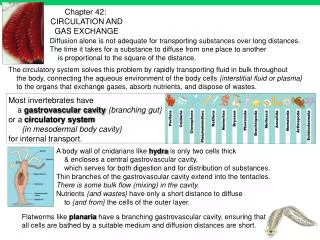
Gastrovascular Cavities • Wide diversity of animals is paralleled by the diversity in circulatory systems. • Simple animals, such as cnidarians • Have a body wall only two cells thick that encloses a gastrovascular cavity • The gastrovascular cavity • Functions in both digestion and distribution of substances throughout the body • http://bcs.whfreeman.com/thelifewire/content/chp32/32020.html
Open and Closed Circulatory Systems Overcome limitations of Diffusion • More complex animals • Have one of two types of circulatory systems: open or closed • Both of these types of systems have three basic components • A circulatory fluid (blood) • A set of tubes (blood vessels) • A muscular pump (the heart)
Heart Hemolymph in sinusessurrounding ograns Ostia Anterior vessel Lateral vessels Tubular heart Figure 42.3a (a) An open circulatory system • In insects, other arthropods, and most molluscs • Blood bathes the organs directly in an open circulatory system https://www.youtube.com/watch?v=vGriV82EyeA
Heart Interstitialfluid Small branch vessels in each organ Dorsal vessel(main heart) Ventral vessels Auxiliary hearts (b) A closed circulatory system Figure 42.3b • In a closed circulatory system • Blood is confined to vessels and is distinct from the interstitial fluid
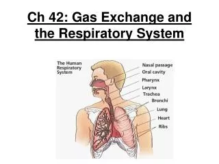
Survey of Vertebrate Circulation • Humans and other vertebrates have a closed circulatory system • Often called the cardiovascular system • Clearly the most advanced/efficient system • Blood flows in a closed cardiovascular system • Consisting of blood vessels and a two- to four-chambered heart
3 Main Types of Blood Vessels: Arteries, Veins, & Capillaries • Arteries carry blood to capillaries • The sites of chemical exchange between the blood and interstitial fluid • Veins • Return blood from capillaries to the heart
Fishes • A fish heart has two main chambers • One ventricle and one atrium • Blood pumped from the ventricle • Travels to the gills, where it picks up O2 and disposes of CO2
Amphibians • Frogs and other amphibians • Have a three-chambered heart, with two atria and one ventricle • The ventricle pumps blood into a forked artery • That splits the ventricle’s output into the pulmocutaneous circuit and the systemic circuit
Reptiles (Except Birds) • Reptiles have double circulation • With a pulmonary circuit (lungs) and a systemic circuit • Turtles, snakes, and lizards • Have a three-chambered heart
Mammals and Birds • In all mammals and birds • The ventricle is completely divided into separate right and left chambers • The left side of the heart pumps and receives only oxygen-rich blood • While the right side receives and pumps only oxygen-poor blood • A powerful four-chambered heart • Was an essential adaptation of the endothermic way of life characteristic of mammals and birds
Gill capillaries Lung and skin capillaries Lung capillaries Lung capillaries AMPHIBIANS REPTILES (EXCEPT BIRDS) MAMMALS AND BIRDS FISHES Right systemicaorta Pulmonarycircuit Artery Pulmocutaneouscircuit Pulmonarycircuit Gillcirculation Heart:ventricle (V) Left Systemicaorta A A A A A A Atrium (A) V V V V V Left Right Left Left Right Right Systemiccirculation Systemic circuit Systemic circuit Vein Systemic capillaries Systemic capillaries Systemic capillaries Systemic capillaries Figure 42.4 • Vertebrate circulatory systems
Double circulation in mammals depends on the anatomy and pumping cycle of the heart • The structure and function of the human circulatory system • Can serve as a model for exploring mammalian circulation in general • Heart valves • Dictate a one-way flow of blood through the heart
Blood begins its flow • With the right ventricle pumping blood to the lungs • In the lungs • The blood loads O2 and unloads CO2
Oxygen-rich blood from the lungs • Enters the heart at the left atrium and is pumped to the body tissues by the left ventricle • Blood returns to the heart • Through the right atrium
Capillaries of head and forelimbs Anterior vena cava Aorta Pulmonary artery Pulmonary artery 9 6 3 3 8 7 Capillaries of right lung Capillaries of left lung 2 4 11 Pulmonary vein Pulmonary vein Left atrium 5 1 Right atrium 10 Left ventricle Right ventricle Aorta Posterior vena cava Capillaries of abdominal organs and hind limbs Figure 42.5 • The mammalian cardiovascular system http://www.sumanasinc.com/webcontent/animations/content/human_heart.html
Pulmonary artery Aorta Pulmonaryveins Pulmonaryartery Anterior vena cava Leftatrium Right atrium Pulmonaryveins Atrioventricularvalve Semilunarvalve Semilunarvalve Atrioventricularvalve Posterior vena cava Right ventricle Figure 42.6 Left ventricle The Mammalian Heart: A Closer Look • A closer look at the mammalian heart • Provides a better understanding of how double circulation works http://highered.mcgraw-hill.com/sites/0072495855/student_view0/chapter22/animation__the_cardiac_cycle__quiz_1_.html
The heart contracts and relaxes • In a rhythmic cycle called the cardiac cycle • The contraction, or pumping, phase of the cycle • Is called systole • The relaxation, or filling, phase of the cycle • Is called diastole
The heart rate, also called the pulse • Is the number of beats per minute • The cardiac output • Is the volume of blood pumped into the systemic circulation per minute • Some cardiac muscle cells are self-excitable • Meaning they contract without any signal from the nervous system
A region of the heart called the sinoatrial (SA) node, or pacemaker • Sets the rate and timing at which all cardiac muscle cells contract • Impulses from the SA node • Travel to the atrioventricular (AV) node • At the AV node, the impulses are delayed • And then travel to the Purkinje fibers that make the ventricles contract http://highered.mcgraw-hill.com/sites/0072495855/student_view0/chapter22/animation__conducting_system_of_the_heart.html
Signals pass to heart apex. Signals spread Throughoutventricles. Pacemaker generates wave of signals to contract. Signals are delayed at AV node. Bundlebranches AV node SA node(pacemaker) Purkinjefibers Heartapex ECG 2 1 3 4 Figure 42.8 • The control of heart rhythm
Structural differences in arteries, veins, and capillaries • Correlate with their different functions • Arteries have thicker walls • To accommodate the high pressure of blood pumped from the heart
Direction of blood flowin vein (toward heart) Valve (open) Skeletal muscle Valve (closed) Figure 42.10 • In the thinner-walled veins • Blood flows back to the heart mainly as a result of muscle action
Blood Pressure • Systolic pressure • Is the pressure in the arteries during ventricular systole • Is the highest pressure in the arteries • Diastolic pressure • Is the pressure in the arteries during diastole • Is lower than systolic pressure
The critical exchange of substances between the blood and interstitial fluid • Takes place across the thin endothelial walls of the capillaries • The lymphatic system • Returns fluid to the body from the capillary beds • Aids in body defense
Blood is a connective tissue with cells suspended in plasma • Blood in the circulatory systems of vertebrates • Is a specialized connective tissue
Blood Composition and Function • Blood plasma is about 90% water • Among its many solutes are • Inorganic salts in the form of dissolved ions, sometimes referred to as electrolytes • Blood consists of several kinds of cells • Suspended in a liquid matrix called plasma • The cellular elements • Occupy about 45% of the volume of blood
Plasma 55% Constituent Major functions Solvent for carrying other substances Water Icons (blood electrolytes Sodium Potassium CalciumMagnesium Chloride Bicarbonate Osmotic balance pH buffering, and regulation of membrane permeability Separatedbloodelements Plasma proteins Albumin Fibringen Immunoglobulins (antibodies) Osmotic balance, pH buffering Clotting Defense Substances transported by blood Nutrients (such as glucose, fatty acids, vitamins) Waste products of metabolism Respiratory gases (O2 and CO2) Hormones Figure 42.15 • The composition of mammalian plasma
Cellular Elements • Suspended in blood plasma are two classes of cells • Red blood cells, which transport oxygen • White blood cells, which function in defense • A third cellular element, platelets • Are fragments of cells that are involved in clotting
Separatedbloodelements Cellular elements 45% Functions Cell type Numberper L (mm3) of blood • The cellular elements of mammalian blood Erythrocytes(red blood cells) Transport oxygenand help transportcarbon dioxide 5–6 million Defense andimmunity Leukocytes(white blood cells) 5,000–10,000 Lymphocyte Basophil Eosinophil Neutrophil Monocyte Platelets 250,000400,000 Blood clotting Figure 42.15
Erythrocytes • Red blood cells, or erythrocytes • Are by far the most numerous blood cells • Transport oxygen throughout the body • The blood contains five major types of white blood cells, or leukocytes • Monocytes, neutrophils, basophils, eosinophils, and lymphocytes, which function in defense by phagocytizing bacteria and debris or by producing antibodies
Pluripotent stem cells(in bone marrow) Lymphoidstem cells Myeloidstem cells Basophils B cells T cells Lymphocytes Eosinophils Neutrophils Erythrocytes Monocytes Platelets Figure 42.16 • Erythrocytes, leukocytes, and platelets all develop from a common source • A single population of cells called pluripotent stem cells in the red marrow of bones
Cardiovascular Disease • Cardiovascular diseases • Are disorders of the heart and the blood vessels • Account for more than half the deaths in the United States
Smooth muscle Connective tissue Plaque Endothelium (a) Normal artery (b) Partly clogged artery 50 µm 250 µm Figure 42.18a, b • One type of cardiovascular disease, atherosclerosis • Is caused by the buildup of cholesterol within arteries
Hypertension, or high blood pressure • Promotes atherosclerosis and increases the risk of heart attack and stroke • A heart attack • Is the death of cardiac muscle tissue resulting from blockage of one or more coronary arteries • A stroke • Is the death of nervous tissue in the brain, usually resulting from rupture or blockage of arteries in the head
Respiratorymedium(air of water) O2 CO2 Respiratorysurface Organismal level Circulatory system Cellular level Energy-richmoleculesfrom food ATP Cellular respiration Figure 42.19 Gas Exchange Occurs Across Specialized Respiratory Surfaces • Generally referred to as Respiration • Do not confuse with cellular respiration which refers energy transformations • Gas exchange supplies oxygen for cellular respiration and disposes of carbon dioxide
Remember key biological concept….lots of body structure have numerous folds to increase surface area for chemical reaction • The part of an animals body where gas exchange occurs is called a respiratory surface • Animals require large, moist respiratory surfaces for the adequate diffusion of respiratory gases • Between their cells and the respiratory medium, either air or water • Rate of diffusion is proportional to surface area across which diffusion occurs and inversely proportional to the square of the distance molecules must move • As a result respiratory surfaces tend to be thin and have large surface areas
Critical Thinking: Who will have more surface area in their respiratory surfaces, ectotherms or endotherms? Explain. Endotherms. While respiratory area depends mainly on the size of the organism and whether it lives in a terrestrial or aquatic environment, endotherms utilize more energy (ATP) and so must do more cellular respiration than a similarly sized ectotherm
Skin as a Respiratory Organ • Some animals use entire outer skin as a respiratory organ • Earthworms and amphibians • Moist skin exchanges gasses by diffusion across entire body • Respiratory surface must remain moist requiring these organisms to live in water or damp places • Organisms usually small (thin or flat)with high surface area to volume ratios
3 Most Common Respiratory Organs • Gills: out-foldings of body surface suspended in water • Respiratory medium= water • The warmer and saltier the water, the less dissolved O2 • Trachea: made up of air tubes that branch throughout the body • Most common respiratory system of terrestrial animals (insects) • Lungs: most familiar respiratory system
(a) Sea star. The gills of a sea star are simple tubular projections of the skin. The hollow core of each gillis an extension of the coelom(body cavity). Gas exchangeoccurs by diffusion across thegill surfaces, and fluid in thecoelom circulates in and out ofthe gills, aiding gas transport. The surfaces of a sea star’s tube feet also function in gas exchange. Gills Coelom Figure 42.20a Tube foot • In some invertebrates • The gills have a simple shape and are distributed over much of the body
Oxygen-poorblood Gill arch Oxygen-richblood Lamella Blood vessel Gill arch 15% 40% 70% 5% Water flow 30% Operculum 100% 60% 90% O2 Blood flowthrough capillariesin lamellaeshowing % O2 Water flowover lamellaeshowing % O2 Gillfilaments Figure 42.21 Countercurrent exchange • The effectiveness of gas exchange in some gills, including those of fishes • Is increased by ventilation and countercurrent flow of blood and water • Ram ventilation • Appendages that “paddle” water past the gills
Advantages & Disadvantages to Air as the Respiratory Medium Advantages of air Disadvantages of air Respiratory surfaces must be large and MOIST Air dries things out • Higher concentration of dissolved oxygen • Oxygen and carbon dioxide diffuse much faster in air than water, so respiratory surfaces have to be ventilated much less vigorously in air than water • When ventilation does occur in terrestrial animals it requires less energy because air is lighter than water and easier to pump
Air sacs Tracheae Spiracle (a) The respiratory system of an insect consists of branched internal tubes that deliver air directly to body cells. Rings of chitin reinforce the largest tubes, called tracheae, keeping them from collapsing. Enlarged portions of tracheae form air sacs near organs that require a large supply of oxygen. Air enters the tracheae through openings called spiracles on the insect’s body surface and passes into smaller tubes called tracheoles. The tracheoles are closed and contain fluid (blue-gray). When the animal is active and is using more O2, most of the fluid is withdrawn into the body. This increases the surface area of air in contact with cells. Figure 42.22a Tracheal Systems in Insects • The tracheal system of insects • Consists of tiny branching tubes that penetrate the body
Body cell Airsac Tracheole Body wall Myofibrils Trachea Air Tracheoles Mitochondria (b) This micrograph shows cross sections of tracheoles in a tiny piece of insect flight muscle (TEM). Each of the numerous mitochondria in the muscle cells lies within about 5 µm of a tracheole. Figure 42.22b 2.5 µm • The tracheal tubes • Supply O2 directly to body cells
With virtually all body cells within a very short distance of respiratory medium, the open circulatory system of insects is not involved in transporting O2 and CO2 For small insects, diffusion through the trachea brings in enough oxygen and removes enough carbon dioxide to support cellular respiration Larger insects with higher energy demands ventilate their tracheal systems with rhythmic body movements that compress and expand the air tubes like bellows
Lungs Unlike the branching tracheal system of insects, lungs are confined to 1 location Because the respiratory surface of a lung is not in direct contact with all other body cells, the gap must be bridged by the circulatory system
Evolution of Lungs • Lungs have evolved in spiders, terrestrial snails, and most vertebrates • Amphibians have small poorly developed lungs due to the gas exchange in their skin • Some have no lungs at all • All mammals rely entirely on lungs • Exceptions • Turtles supplement lung breathing with gas exchange in the mouth and anus pew • lungfish, fish with lungs to live for short periods of time outside of water or in oxygen poor water
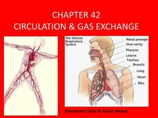
Branch from thepulmonaryartery(oxygen-poor blood) Branch from the pulmonary vein (oxygen-rich blood) Terminal bronchiole Nasalcavity Alveoli Pharynx Left lung Esophagus Larynx 50 µm Trachea 50 µm Right lung Bronchus Bronchiole Colorized SEM SEM Diaphragm Heart Figure 42.23 Mammalian Respiratory Systems: A Closer Look • A system of branching ducts • Conveys air to the lungs http://bio-animations.blogspot.com/2008/04/human-body-circulatory-system.html