Download
1 / 38
500 likes | 1.31k Vues
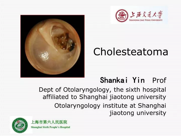
Cholesteatoma. Shankai Yin Prof Dept of Otolaryngology, the sixth hospital affiliated to Shanghai jiaotong university Otolaryngology institute at Shanghai jiaotong university. Epidemiology. Exact prevalence is unknown Incidence estimated between 3 and 12.6 per 100,000. Classification.

E N D
Cholesteatoma Shankai Yin Prof Dept of Otolaryngology, the sixth hospital affiliated to Shanghai jiaotong university Otolaryngology institute at Shanghai jiaotong university
Epidemiology • Exact prevalence is unknown • Incidence estimated between 3 and 12.6 per 100,000
Classification • Congenital • Acquired • Primary acquired (retraction pocket) • Secondary acquired
Pathogenesis • Congenital • Arise from embryonal rests of epithelial cells • Location (petrous pyramid, mastoid and middle ear cleft) • Levenson criteria • White mass medial to normal TM • Normal pars flaccida and tensa • No history of otorrhea or perforations • No prior otologic procedures • Prior bouts of otitis media not grounds for exclusion
Theories “Acquired” inclusion theory - Tos Epidermal rest theory - Teed Michael
Primary acquired • Eustachian tube dysfunction • Poor aeration of the epitympanic space • Retraction of the pars flaccida • Normal migratory pattern altered • Accumulation of keratin, enlargement of sac
Secondary acquired • Implantation – surgery, foreign body, blast injury • Metaplasia – transformation of cuboidal epithelium to squamous epithelium from chronic infection • Invasion/Migration – medial migration along permanent perforation of TM • Papillary ingrowth – intact pars flaccida, inflammation in Prussack’s space, break in the basal membrane, cords of epithelium migrate inward
Clinical manifestations • Common • Painless otorrhea • Refractory/recurrent ear infections • Conductive hearing loss • Uncommon • Vertigo/Sensorineural • Facial nerve paralysis • CNS infections • Brain herniation/CSF leak • Pneumocephalus
Diagnosis • history • Physical Examination • Otomicroscopy • Posterosuperior retraction pocket with squam • Granulation from diseased bone • Aural polyps • Pneumatic otoscopy – positive fistula response suggests erosion into labyrinth • Cultures should be obtained in infected ears
Audiology • usually conductive loss, may vary greatly; confirm with tuning forks • Imaging • CT temporal bone – definitely obtain for revision cases, complications of chronic suppurative otitis media, sensorineural hearing loss, vestibular symptoms, other complications of cholesteatoma
Imaging • Purpose • Diagnosis • Determining extent • Risk assessment • Modalities • Plain film • Computed tomography scans • Magnetic Resonance imaging
Goals of CT Imaging • Middle ear ventilation • Ossicular destruction • Epitympanum access • Mastoid cortex • Tegmen integrity • Labyrinth involvement • Facial nerve involvement • Surgical changes
CT disadvantages • Granulation tissue vs. cholesteatoma • Specific soft tissue problems • Dural involvement • Abscess • Brain herniation • Labyrinth involvement • Sigmoid sinus thrombosis • MRI needed
MR Imaging • Hypointense on T1 • Isointense to brain • Intermediate on T2 • Nonenhancing • Granulation tissue does enhance • Recurrence detection • Lesions >2mm • 90% sensitive, 100% specificity
T2 Delayed contrast T1
Differential Diagnosis • Chronic serous otitis media • Jugulotympanic paragangliomas • Cholesterol granulomas • Neurofibromas • Hemangiomas • Arachnoid cyst • Jugular bulb anomalies • Tympanosclerosis • encephalocele
Treatment Create a “dry and safe” ear
Non-surgical • Treat the Infection – Floxin Otic Drops • Decrease the inflammation – Topical steroids • Debridement of the external canal
Surgical • Atticotomy • Radical Mastoidectomy • Bondy Modified Radical (Canal wall down) mastoidectomy • Tympanoplasty and canal wall up mastoidectomy
Prognosis • Residual or recurrent cholesteatoma over 5 years – 15 to 40% • Reported to be up to 67% in the pediatric population • Close follow - up • Regular examinations needed - 6 months
Complications • Dural tear - CSF leak • Fistula of the horizontal semicircular canal (vertigo) – Up to 10% • Facial nerve injury • Injury to the sigmoid sinus / jugular bulb • Otitic Hydrocephalus • Hearing loss • 30% have conductive loss pre-operatively • Postoperatively, an additional 30% have worsening or onset of hearing loss due to extent of disease • Infection – Meningitis, Abscess, lateral sinus thrombosis – Up to 1%
Predisposing factors • Virulent organisms • Cholesteatoma and bone erosion • Presence of a congenital dehiscence (e.g.dehiscent facial canal) or a preformed pathway (e.g. skull base fracture) • Obstruction of drainage e.g. by a polyp. • Low resistance of the patient
Pathways of infection • The commonest way for extension of infection is by bone erosion due to a cholesteatoma. • Vascular extension (retrograde thrombophlebitis). • Extension along preformed pathways as – Congenital dehiscences, fracture lines, round window membrane, the labyrinth, – Dehiscences due to previous surgery.
Classification • Cranial complications • Extra-cranial complications • Intra-cranial complications
Cranial complications • Acute mastoiditis and mastoid abscesses (most common complication). • Petrositis. • Labyrinthitis. • Facial paralysis. • Osteomyelitis of the temporal bone
Extra-cranial complications • External otitis • Cervical lymphadenitis • Retropharyngeal • Parapharyngeal abscesses
Intracranial complications • Extradural abscess (commonest intracranial complication). • Subdural abscess. • Meningitis. • Brain abscess: • Temporal lobe abscess. • Cerebellar abscess. • Lateral sinus thrombosis. • Otitic hydrocephalus.
Potentially life threatening • Suppurative otorrhea, chronic headache, pain, fever – impending intracranial complication • Mental status changes, nuchal rigidity, cranial neuropathies require neurosurgical consult