Download
1 / 33
410 likes | 866 Vues
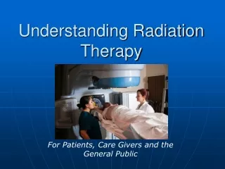
Understanding radiation units L02. IAEA Training Course on Radiation Protection for Doctors (non-radiologists, non-cardiologists) using Fluoroscopy. Educational Objectives. How radiation dose can and should be expressed? Radiation quantities useful for dose to doctors that use fluoroscopy

E N D
Understanding radiation unitsL02 IAEA Training Course on Radiation Protection for Doctors (non-radiologists, non-cardiologists) using Fluoroscopy
Educational Objectives • How radiation dose can and should be expressed? • Radiation quantities useful for dose to doctors that use fluoroscopy • Radiation quantities useful for patient doses from fluoroscopic procedures • Individual monitoring • The inverse square law IAEA Training Course on Radiation Protection for Doctors (non-radiologists, non-cardiologists) using Fluoroscopy L02. Understanding radiation units
500 mg of anti-inflammatory drug. • Dose outside (in drug) is same as dose inside the patient’s body Is the situation same with radiation? IAEA Training Course on Radiation Protection for Doctors (non-radiologists, non-cardiologists) using Fluoroscopy L02. Understanding radiation units
Not so in case of radiation. • It depends upon the absorption on body surface (in air) Absorbed dose In tissue • Difficult (rather not possible) to measure dose inside the body • Solution: measure dose in air, then estimate/calculate dose in tissue IAEA Training Course on Radiation Protection for Doctors (non-radiologists, non-cardiologists) using Fluoroscopy L02. Understanding radiation units
Examples of radiation measures Watt Attenuation by paper or metal object IAEA Training Course on Radiation Protection for Doctors (non-radiologists, non-cardiologists) using Fluoroscopy L02. Understanding radiation units
Why so many quantities? • 1000 W heater giving heat (IR radiation)- unit is infra red power which is related with emission intensity • Heat perceived by the person will vary with so many factors:distance, clothing, temperature in room… • If one has to go a step ahead, from perception of heat to heat absorbed, it becomes a highly complicated issue • This is the case with X rays– They can’t even be perceived IAEA Training Course on Radiation Protection for Doctors (non-radiologists, non-cardiologists) using Fluoroscopy L02. Understanding radiation units
Whole body dose in mSv • Organ dose in mGy • Dose falling on the body in mGy How to define mGy, mSv? IAEA Training Course on Radiation Protection for Doctors (non-radiologists, non-cardiologists) using Fluoroscopy L02. Understanding radiation units
mGy Equivalent dose to whole body (mSv) mGy mGy mGy x wt=mSv Effective dose (mSv) IAEA Training Course on Radiation Protection for Doctors (non-radiologists, non-cardiologists) using Fluoroscopy L02. Understanding radiation units
Understanding of the Radiation Units • How much radiation do we get from natural sources? • Around 1-3 mSv (which dose is this?) • How much radiation does a patient get from a chest radiograph? • around 0.02 mSv (effective dose) • Staff dose obtained from radiation badge? • Effective dose https://rpop.iaea.org/RPOP/RPoP/Content/InformationFor/Patients/information-public/index.htm# IAEA Training Course on Radiation Protection for Doctors (non-radiologists, non-cardiologists) using Fluoroscopy L02. Understanding radiation units
Absorption & biological effects • Different tissues have different sensitivities to ionizing radiation • Same amount of radiation will have different effect in different tissues • The higher the weighting factor, the more sensitive the tissue IAEA Training Course on Radiation Protection for Doctors (non-radiologists, non-cardiologists) using Fluoroscopy L02. Understanding radiation units
Relative Dose Received With modern systems eff dose for chest =0.02 mSv mSv .05 Arm, head,ankle & foot (1) 0.15 Head & Neck (3) Head CT (10) 0.49 Thoracic Spine (18) 0.92 Mammography, Cystography (20) 1.0 Pelvis (24) 1.22 Abdomen, Hip, Upper & lower femur (28) 1.4 Ba Swallow (30) 1.5 Obsteric abdomen (34) 1.7 Lumbo-sacral area (43) 2.15 Cholangiography (52) 2.59 Lumber Myelography (60) 3.0 Lower abdomen CT male (72) 3.61 Upper Abdomen CT (73) Ba Meal (76) 3.67 Angio-head, Angio-peripheral (80) 3.8 Urography (87) 4.0 Angio-abdominal (120) 4.36 Chest CT (136) 6.0 Lower Abd. CT fem. (142) 6.8 Ba enema (154) 7.13 Lymphan. (180) 7.69 9.0 0 50 100 150 200 Typical effective doses from radiological examinations (expressed as equal number of chest X rays) number of chest X rays IAEA Training Course on Radiation Protection for Doctors (non-radiologists, non-cardiologists) using Fluoroscopy L02. Understanding radiation units
Staff exposure- which Units? • Effective dose-overall risk, personal dose equivalent Hp(10) • Skin dose-injuries (fingers, legs) • Organ dose-cataract, thyroid, gonads IAEA Training Course on Radiation Protection for Doctors (non-radiologists, non-cardiologists) using Fluoroscopy L02. Understanding radiation units
Limits on Occupational Doses (ICRP)* 1In a recent statement ICRP (Statement on Tissue Reactions Approved by the Commission on April 21, 2011) the ICRP proposed a limit of 20 mSv/yr, averaged over 5 years, not exceeding 50 mSv at any single year. *Please follow the recommendations as prescribed by your national authority IAEA Training Course on Radiation Protection for Doctors (non-radiologists, non-cardiologists) using Fluoroscopy L02. Understanding radiation units
Patient doses • Peak skin dose • Cumulative air kerma • kerma Area Product (KAP or DAP) IAEA Training Course on Radiation Protection for Doctors (non-radiologists, non-cardiologists) using Fluoroscopy L02. Understanding radiation units
Peak skin dose 1 2 3 Example of dose distribution in a Coronary angiography procedure shown using self-darkening film. Dose is determined by measuring darkening and relating it to the films Characteristic curve After scanning the Film and using the dose curve, a dose map can Be created IAEA Training Course on Radiation Protection for Doctors (non-radiologists, non-cardiologists) using Fluoroscopy L02. Understanding radiation units
Commonly used patient dose descriptors • Radiography and fluoroscopy: • Entrance Surface Dose (ESD) [mGy] • by measurements (TLD or film) • calculated by tube output factors and exposure geometry • Dose Area Product (DAP) [Gy-cm²] • by real-time measurement with DAP meter • Computed Tomography: • Computed Tomography Dose Index (CTDI) [mGy] • Dose length product (DLP) [mGycm] IAEA Training Course on Radiation Protection for Doctors (non-radiologists, non-cardiologists) using Fluoroscopy L02. Understanding radiation units
Dose Area Product (DAP) or air-Kerma Area Product (KAP)* The dose-area product (DAP) quantity is defined as the dose in air in a plane, integrated over the area of interest. DAP is expressed in Gy-cm². DAP is automatically measured in real-time on most recent fluoroscopy systems *preferred term IAEA Training Course on Radiation Protection for Doctors (non-radiologists, non-cardiologists) using Fluoroscopy L02. Understanding radiation units
d1=1 Area = 1Dose = 1 d2=2 Area = 4Dose = 1/4 FluoroscopyKerma- area product • KAP = D x Area the SI unit of KAP is the Gy.cm2 Current name KAP KAP is independent of distance from the source IAEA Training Course on Radiation Protection for Doctors (non-radiologists, non-cardiologists) using Fluoroscopy L02. Understanding radiation units
KAP Real-time KAP values displayed on monitor IAEA Training Course on Radiation Protection for Doctors (non-radiologists, non-cardiologists) using Fluoroscopy L02. Understanding radiation units
TLD or film Entrance surface dose (ESD) • The ESD is measured on the surface of a patient or a phantom • It can also be calculated by using the tube output and geometrical description of the procedure • It can be used as a metric to estimate risk of deterministic skin effects IAEA Training Course on Radiation Protection for Doctors (non-radiologists, non-cardiologists) using Fluoroscopy L02. Understanding radiation units
Using KAP and ESD in practice Example from vascular surgery procedure Endovascular aneurysm repair (EVAR) Repair of abdominal aortic aneurism (AAA) R Weerakkody et al, BJS 2008 IAEA Training Course on Radiation Protection for Doctors (non-radiologists, non-cardiologists) using Fluoroscopy L02. Understanding radiation units
Using KAP and ESD in practice Example from orthopaedic surgery procedure Exposure factors for four projections commonly used in orthopaedic surgery From N Theocharopoulos et al; Journal of Bone and Joint Surgery 2003 IAEA Training Course on Radiation Protection for Doctors (non-radiologists, non-cardiologists) using Fluoroscopy L02. Understanding radiation units
Occupational exposure Individual monitoring and exposure assessment IAEA Training Course on Radiation Protection for Doctors (non-radiologists, non-cardiologists) using Fluoroscopy L02. Understanding radiation units
Thank you IAEA Training Course on Radiation Protection for Doctors (non-radiologists, non-cardiologists) using Fluoroscopy L02. Understanding radiation units
Extra Slides IAEA Training Course on Radiation Protection for Doctors (non-radiologists, non-cardiologists) using Fluoroscopy L02. understanding radiation units
Kerma Kinetic Energy Released in Matter IAEA Training Course on Radiation Protection for Doctors (non-radiologists, non-cardiologists) using Fluoroscopy L02. Understanding radiation units 27
KermaKinetic Energy Released in Matter • KERMA is defined as the sum of the initial kinetic energies of all electrons released by X ray photons per unit mass. • It is no longer recommended to use the term “absorbed dose in air” (ICRU 2005) when speaking of ionization in air. • KERMA is a precursor to absorbed dose. IAEA Training Course on Radiation Protection for Doctors (non-radiologists, non-cardiologists) using Fluoroscopy L02. Understanding radiation units
Absorbed dose, D and KERMA • The KERMA(kinetic energy released in a mass) The SI unit of kerma is the joule per kilogram (J/kg), termed gray (Gy). IAEA Training Course on Radiation Protection for Doctors (non-radiologists, non-cardiologists) using Fluoroscopy L02. Understanding radiation units
Dose quantities and radiation units Absorbed dose The absorbed dose D, is the energy absorbed per unit mass in a medium SI unit of D is the gray [Gy] Entrance surface dose includes the scatter from the patientESD D * 1.4 IAEA Training Course on Radiation Protection for Doctors (non-radiologists, non-cardiologists) using Fluoroscopy L02. Understanding radiation units
Absorbed Dose The absorbed dose D, is the energy absorbed per unit mass in a medium SI unit of D is the gray [Gy] Entrance surface dose includes the back-scatter from the patientESD D * 1.4 IAEA Training Course on Radiation Protection for Doctors (non-radiologists, non-cardiologists) using Fluoroscopy L02. Understanding radiation units 31
Mean absorbed dose in a tissue or organ • The mean absorbed dose in a tissue or organ DT is the energy deposited in the organ divided by the mass of that organ. IAEA Training Course on Radiation Protection for Doctors (non-radiologists, non-cardiologists) using Fluoroscopy L02. Understanding radiation units 32
Equivalent dose (H) The equivalent dose H is the average absorbed dose in an organ multiplied by a dimensionless radiation weighting factor, wR which expresses the biological effectiveness of a given type of radiation H = D * wR the SI unit of H is the sievert [Sv] For X rays is wR=1 Note: This unit H, is not often used in radiological imaging and is often confused with Effective dose IAEA Training Course on Radiation Protection for Doctors (non-radiologists, non-cardiologists) using Fluoroscopy L02. Understanding radiation units
Effective dose, E The equivalent doses to organs and tissues weighted by the relative wT are summed over the whole body to give the effective dose E E = sum of (equivalent doses xwT) wT : weighting factor for organ or tissue T This unit is OFTEN used in radiological imaging and can be used to compare dose efficiency of competing imaging examinations IAEA Training Course on Radiation Protection for Doctors (non-radiologists, non-cardiologists) using Fluoroscopy L02. Understanding radiation units